Overview
Researchers at Yonsei University and Jungmok Seo (then at the Korea Institute of Science and Technology, KIST, and a corresponding author), who now leads the BLISS Lab at Yonsei University, developed a droplet manipulation system in which a vacuum tip deforms a stretchable superamphiphobic (SPO) polydimethylsiloxane (PDMS) substrate from below and superamphiphilic (SPI) spots retain small aliquots for colorimetric assays. Water aliquots of 0.05 to 1 μL were dispensed on 1 to 2 mm spots, and glucose, uric acid and lactate were read from a single droplet.
Background
Droplet-based microfluidics handles small samples without channel networks, pumps or tubing, but actuation by electrowetting, magnetic force, light or acoustic waves often requires additives such as electrolytes or magnetizable particles, and electrowetting requires a closed system with high applied voltage. An earlier deformable superhydrophobic platform from the same authors could move water droplets in any direction but could not dispense small amounts of liquid from the sample. To the authors' knowledge, no droplet-based system had allowed full-range manipulation of low-surface-tension and viscous biofluidic droplets such as blood.
Design and approach
The SPO layer is made by spraying an adhesive and then fluorinated silica nanoparticles onto a thin PDMS sheet (about 340 μm), which forms re-entrant micro- and nanostructures with low surface energy. Oxygen plasma through a shadow mask (100 W for 45 s) decomposes the fluoroalkyl silane in exposed regions and converts them to SPI spots. A vacuum tip beneath the suspended sheet creates a moving local dimple; the droplet follows the dimple over the weakly adhesive SPO region, and when it crosses an SPI spot the spot pins part of the liquid. A daughter droplet separates when the gravitational force on the parent droplet exceeds the Furmidge adhesion force of the spot, so the minimum parent mass and the dispensed volume depend on spot diameter and surface tension.
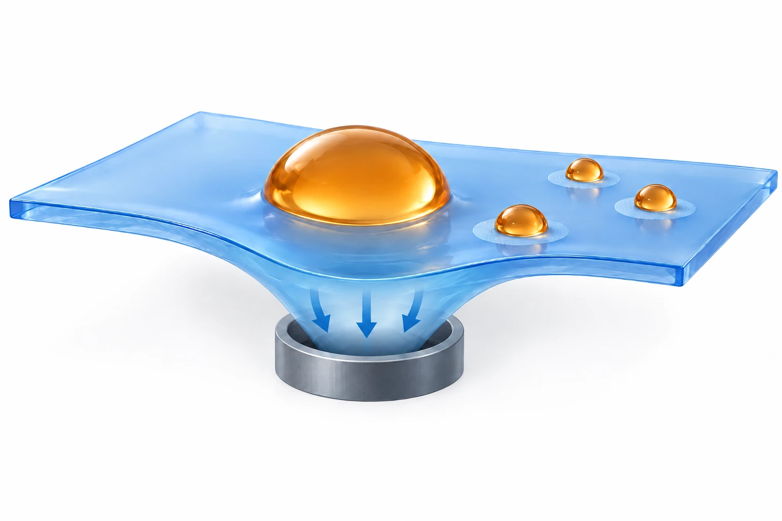
AI-generated, not-to-scale concept showing how a vacuum tip deforms a substrate to guide droplets while wettable patterns retain some liquid. Arrows show substrate deformation and circles mark wettable regions.
Generated for this library to illustrate the design; it is not a figure from the paper and does not show measured data.
How it was evaluated
Wettability was measured for water, ethylene glycol (EG), heparinized blood (0.75 U/mL heparin), dimethyl sulfoxide and hydrogels, and contact and sliding angles of EG droplets were measured under uniaxial strain and repeated stretching. Droplet guiding used a 10 mm vacuum tip at 20 kPa moved at 3 mm/s, with SPI spots of 2 mm diameter and 15 mm spacing. Dispensing was characterized as a function of spot diameter for water and EG. For the assays, 1 μL of enzyme and chromogen reagent was dispensed and dried on each spot, followed by 1 μL of sample; glucose, uric acid and lactate were read by scanning color intensity, and glucose was measured in samples from healthy and diabetic ICR mice and compared with a commercial glucometer.
Key results
The SPO surface showed contact angles above 150° and sliding angles below 10° for all tested liquids, while SPI regions showed a near-zero contact angle. Repellency was retained for liquids with surface tension above 33 mN/m, which is lower than that of blood, plasma, sweat and urine (46 to 72 mN/m). The coating kept its liquid repellency at 100% uniaxial strain and showed no defects or delamination after 1000 stretch and release cycles at 100% strain, with unchanged contact and sliding angles.
The adhesion force of the SPO surface was calculated as 2.6 mN, which implies that water droplets larger than about 3 μL can follow the dimple. A 200 μL EG droplet traveled with the tip and left a few microliters on each SPI spot along its path. As spots increased from 1 to 2 mm in diameter, daughter droplets of 0.05 to 1 μL (water) and 0.4 to 1.4 μL (EG) were dispensed, and water dispensing worked on spots as small as 0.6 mm. A red-dyed spot kept its wettability for a second droplet, and successive rhodamine B and malachite green deposits on one spot gave both absorption peaks (557 and 627 nm), suggesting that sequentially dispensed liquids can mix and react on a spot.
Color intensity was linear over 0 to 20 mM glucose, 0.25 to 4 mM uric acid and 0 to 20 mM lactate, and the assay substrate kept stable wettability and colorimetric response after 2 weeks of storage. In a 3 × 3 array with one reagent per column, three analyte mixtures were dispensed from single droplets, and the measured concentrations matched the predesigned values within a 20% error, which the authors relate to the FDA standard. Glucose was 13.3 mM in the diabetic mouse sample and 7.5 mM in the healthy one, against a diabetes threshold of 13 mM, and a commercial glucometer gave similar results.
Open questions
The measured minimum parent mass for dispensing was smaller than the calculated value, and the gap grew with spot diameter, which the authors attribute to nonuniform adhesion caused by deformation of the droplet meniscus. Water was not dispensed on spots smaller than 0.6 mm because their adhesion force was negligible relative to the surface tension of water. The mouse samples are described as plasma in the Abstract and as sera in the Methods.
Related research
Evaluation of the Droplet-Microarray Platform for High-Throughput Screening of Suspension Cells.
DOI: 10.1177/2211068216677204 ↗
The related abstract describes wetting-patterned droplet arrays for parallel treatment and imaging of suspension cells.
Abstract-based introductionRESEARCH NEWS
Research in the news
체액 한 방울만으로 몸속 질병 진단한다 ↗
국내 연구진, 체액 한 방울로 질병 진단하는 바이오센서 기술 개발 ↗
KIST·연세대 연구팀, 혈액검사 혈액량 100분의 1로 줄이는 기술 개발 ↗
혈액 한 방울로 질병진단 가능해진다 ↗
[News] “미세 액적 제어 시스템” 언론 보도 ↗
Unverified candidates and access limits (3)
Unverified candidate · 바이오센서용 나노코팅기판 개발…소량 시료로 정밀측정 가능 ↗
Unverified candidate · '체액 한 방울'로 다양한 질병 동시 진단 ↗
Unverified candidate · 국내 연구진, 체액 한 방울로 질병 진단 기술 개발 ↗
Sources used in this summary
This summary is based on the publisher PDF of the article (ACS Nano 2018, 12, 932-941; 10 pages, local copy), including the abstract, main text, figure legends, Table 1, Methods and author information; the Supporting Information and movies were not reviewed, and values shown only in figure panels were not used.