Overview
This review, written by the BLISS Lab (Jungmok Seo) at Yonsei University, examines how implantable biosensors and medical implants can be protected from the foreign body response (FBR), the nonspecific immune reaction that the authors identify as the main cause of implant complications. It organizes the field into three groups of strategies: materials and mechanics, drug delivery, and biomimetic surfaces. Korean national statistics cited in the review put complication surgeries after total knee replacement and pacemaker implantation at approximately 3.2%.
Background
Implant numbers have risen with population aging, and complication surgeries related to artificial implants in Korea have increased since 2008. The FBR proceeds through protein adhesion, immune cell adhesion and fibrous capsule formation; acute inflammation lasts from a few hours to several days and, when a large implant cannot be removed by phagocytosis, gives way to chronic inflammation, foreign body giant cells and a fibrous capsule. For implantable biosensors, adsorbed proteins and cells raise electrode impedance, which reduces sensitivity and lifetime and makes recalibration necessary. The authors also note that a material found safe in one implant application cannot be assumed safe in all others.
How the review is organized
The review first describes the inflammatory cascade at the implant surface and then discusses interventions in three categories. Materials and mechanics covers flexible substrates that reduce mechanical mismatch with soft tissue, biocompatible natural and synthetic coatings, and surface micropatterning. Drug delivery covers anti-inflammatory and anti-fibrotic drugs, carriers that control where and when drugs are released, and implanted actuators. Biomimetic surfaces covers lotus-inspired superhydrophobic surfaces and Nepenthes-inspired slippery liquid-infused porous surfaces (SLIPS) with their medical applications.
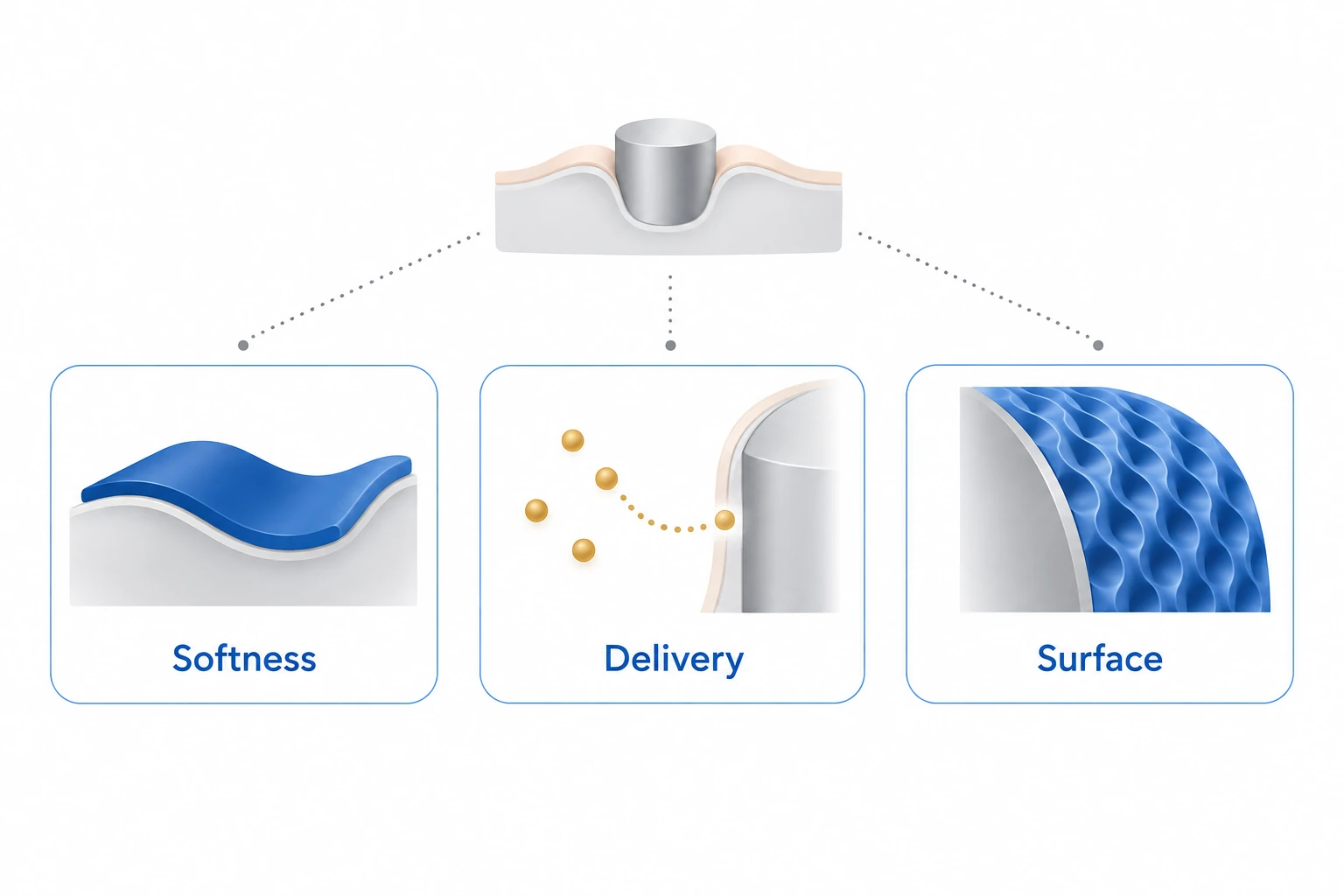
AI-generated concept showing the three strategy categories in the review abstract: soft materials, drug delivery and biomimetic surfaces for addressing foreign-body responses.
Generated for this library to illustrate the design; it is not a figure from the paper and does not show measured data.
Scope of the review
Each strategy is discussed through representative studies and figures reproduced from them: neural probes stiffened temporarily for insertion (Figure 2a), a thermoresponsive double network nanocomposite (DNNC) hydrogel membrane and poly(acrylic acid) (PAA) micropatterned poly(dimethylsiloxane) (PDMS) implants (Figure 2b, c), pirfenidone, polymeric nanohydrogels and a magnetically driven insulin pump (Figure 3), and SLIPS, a tethered-liquid perfluorocarbon (TLP) blood-contact surface and a lubricant-infused surface (LIS) for electrosurgical instruments (Figure 4). For several strategies, the review also notes what remains unresolved, such as signal loss in flexible probes, the lack of effect of anti-fibrotic agents on early FBR, and reduced pump output after fibrous capsule formation.
Main points of the review
Flexible neural probes reduce damage from micromotion but cannot penetrate the dura mater on their own. Stiffening coatings of metal or silicon are not biodegradable, whereas polyethylene glycol (PEG), carboxymethylcellulose and silk-I coatings dissolve after insertion and return the probe to its flexible state. In a cited partially flexible probe solidified with sucrose gel, the probe pierced 0.6% agarose without buckling, reached its intended 3.6-mm depth in the mouse brain, and produced a wound approximately five times smaller than a rigid probe. The review notes that such probes still lose signal because the immune response is not completely suppressed.
Among coatings, Nafion membranes on glucose sensors lost efficiency through mineralization, and polyurethane modified with silicone and polyethylene oxide (PU-S-PEO) reduced inflammation in rats after 7 weeks. The DNNC hydrogel membrane, which deswells and reswells with local temperature, recruited fewer macrophages than PEG diacrylate (PEG-DA) at 7 and 30 days, and its fibrous capsule was up to 30 μm thick at 30 days. PAA micropatterns grafted on PDMS reduced macrophage recruitment at 2 weeks and capsule thickness over 2, 4 and 8 weeks in rats, although the cited study did not establish the mechanism.
Glucocorticoids, with dexamethasone the most frequently used, act on the inflammatory reaction; anti-fibrotic agents such as pirfenidone act on capsule formation but not on early protein adhesion and acute inflammation; and gene silencing remains at an early stage. In one cited study, small interfering RNA (siRNA) against the mammalian target of rapamycin (mTOR) reduced fibroblast proliferation and collagen type I (COL1A1) expression in vitro but did not significantly change capsule thickness in vivo. Poly(methacrylic acid) (PMAA) nanohydrogels carried hydrophobic paclitaxel and hydrophilic doxorubicin together with pH-triggered release. In a magnetically driven insulin pump, plasma insulin from the constant-dose pump fell after fibrous capsules formed, in contrast to the pump that doubled its dose after 11 days.
Superhydrophobic surfaces are vulnerable to pressure and impact and do not repel low-surface-tension physiological fluids. SLIPS repels blood without staining, retains its sliding angle up to approximately 700 atm, and self-heals as lubricant wicks into damaged areas. A TLP surface reduced protein and platelet adhesion and clot formation compared with an uncoated control surface, and a stainless-steel LIS with octadecyltrichlorosilane and silicone oil reduced soft tissue adhesion to electrosurgical instruments. The review identifies complex fabrication, difficulty with arbitrary shapes and limited in vivo biocompatibility data as obstacles for these surfaces.
Open questions
The authors conclude that material strategies reduce protein and cell adhesion and micromotion damage but cannot completely prevent the FBR, that non-specific drug delivery can cause side effects in adjacent tissues while capsule formation remains unsolved, and that biomimetic surfaces lose their properties over the long term. They propose integrating the strategies through collaboration between bioengineers and clinicians, with validation in in vivo and clinical studies.
Related research
Foreign body reaction to biomaterials
DOI: 10.1016/j.smim.2007.11.004 ↗
Anderson and colleagues describe the foreign body reaction as the end-stage inflammatory and wound-healing response to an implant, involving macrophages and foreign body giant cells at the material surface; this review builds on that sequence to compare intervention strategies.
Abstract-based introductionSources used in this summary
This summary is based on the published PDF of the review (15 pages, including all sections and figure legends); the primary studies it cites were not read.