Overview
This study, co-authored by Jungmok Seo (then at the Nanobio Device Laboratory, School of Electrical and Electronic Engineering, Yonsei University), who now leads the BLISS Lab at Yonsei University, grew few-layer graphene directly on silver (Ag) nanoparticles using poly(methyl methacrylate) (PMMA) as a solid carbon source to make surface-enhanced Raman scattering (SERS) substrates that resist oxidation. After annealing in air, the 1363 cm−1 peak of rhodamine 6G (R6G) fell from 36304 to 113 on bare Ag nanoparticles but only to 24436 on the graphene-encapsulated particles.
Background
Ag nanostructures give strong SERS enhancement, but oxidation of Ag under ambient conditions changes their morphology and chemistry and reduces the Raman signal. Protective layers of TiO2, SiO2, Al2O3 or amorphous carbon have been explored, but thick, pinhole-free layers generally disrupt the near-field interaction between the metal and the analyte molecules and can significantly reduce the SERS intensity. Graphene is a thinner barrier, but transferred graphene or assembled graphene oxide (GO) complicates processing, and growing graphene shells by chemical vapour deposition (CVD) at high temperature makes the size and assembly of the metal nanoparticles hard to control.
Design and approach
Ag nanoparticles with an average diameter of 82 nm were synthesized by the polyol process, mixed with a 2 wt% PMMA solution, drop-cast on SiO2 substrates and dried at 110 °C. Heating in 20 sccm H2 and 70 sccm Ar at 800 °C for 15 min converts the PMMA, by pyrolysis above 400 °C, into an amorphous carbon (a-C) layer that keeps the particles from agglomerating, and Ag then catalyses the conversion of this layer into few-layer graphene. The graphene shell is intended to act as an impermeable barrier to oxygen while remaining thin enough for SERS.
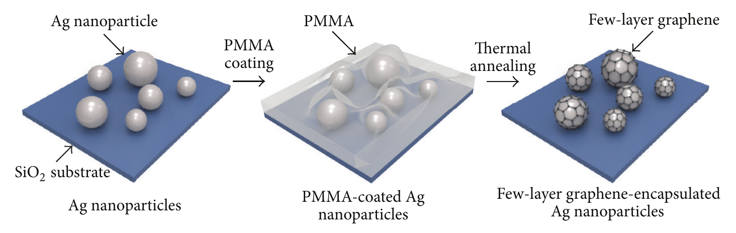
The schematic shows three fabrication stages for forming few-layer graphene on silver nanoparticles using PMMA as the carbon source.
Jaehong Lee et al. (2015). “Highly Stable Surface‐Enhanced Raman Spectroscopy Substrates Using Few‐Layer Graphene on Silver Nanoparticles”. Figure 1. DOI: 10.1155/2015/975043. Creative Commons Attribution; license version is not specified in the source PDF. Cropped to the complete figure from the source PDF; figure content is unchanged.
Source figure and caption ↗ · DOI: 10.1155/2015/975043 ↗ · Creative Commons Attribution; version not specified in the read PDF ↗
How it was evaluated
Particle morphology was compared by scanning electron microscopy (SEM) for the PMMA route and for a methane (CH4) gas route, and the graphene shell was characterized by Raman spectroscopy and high-resolution transmission electron microscopy (HR-TEM). Oxidation resistance was tested by annealing bare and graphene-encapsulated Ag nanoparticles at 150 °C and 200 °C for 2 h under ambient conditions and measuring X-ray diffraction (XRD). SERS performance was measured with R6G (10−6 M, 12 h immersion) under a 633 nm He-Ne laser before and after annealing, and the enhancement factor was calculated for three samples relative to bare silicon at the 1183 cm−1 peak.
Key results
With PMMA as the carbon source, the Ag nanoparticles showed no significant morphological change after growth at 800 °C, whereas the CH4 route produced highly agglomerated, clustered particles. Raman spectra showed overlapping D and G peaks near 1360 and 1590 cm−1 and a broad band at 2300–3200 cm−1; the authors attribute the strong D peak to very small graphene domains on the highly curved particle surfaces, and the D + G and 2D′ components to strained, nonplanar graphene. HR-TEM showed a spacing of about 0.35 nm between coating layers, consistent with the interlayer distance of graphite.
XRD of bare Ag nanoparticles showed the face-centred cubic Ag (111), (200) and (220) peaks at 37.5°, 43.7° and 64.1°, and after annealing in air an Ag2O (111) peak appeared at 32.7° and grew with annealing temperature. For graphene-encapsulated nanoparticles, no Ag oxidation peak was detected after the same annealing.
On bare Ag nanoparticles, the main R6G peaks decreased sharply as the annealing temperature increased, and the 1363 cm−1 peak fell by a factor of 0.003, from 36304 to 113. On graphene-encapsulated nanoparticles, the same peak changed from 36304 to 24436 after annealing. The enhancement factors of three graphene-encapsulated samples were 682, 902 and 697, which the authors take to indicate comparable reproducibility.
Related research
Shell-isolated nanoparticle-enhanced Raman spectroscopy
The related SHINERS paper’s abstract uses silica- or alumina-coated gold particles to limit aggregation and direct sample contact. It offers a comparison of protective coatings and optical access.
Abstract-based introductionRESEARCH NEWS
Research in the news
Sources used in this summary
This summary is based on the publisher PDF of the open-access article, including the experiment section and Figure 1–6 legends; numerical values shown only inside figure panels were not used.