Overview
This study, developed by the BLISS Lab (Jungmok Seo) at Yonsei University with collaborators in the Departments of Biotechnology and of Biomaterials Science and Engineering at Yonsei University and at Seoul National University Hospital and College of Medicine, introduces L-VIP, a stretchable antifouling coating in which a lubricant is infused into a bilayer of the adhesive organosilicon polymer V4D4 and the perfluoropolymer FOMA. The coating kept water contact angles of about 120° and sliding angles below 8° up to 200% strain, showed no cracks after 2000 cycles at 150% strain, and reduced macrophage accumulation and collagen encapsulation around polyvinyl alcohol (PVA) hydrogels implanted subcutaneously in mice.
Background
Implanted bioelectronics are deformed continuously by breathing, circulation and movement, and nonspecific adhesion of proteins, cells and bacteria can trigger foreign body reactions (FBR), fibrotic encapsulation and infection. Hydrophilic polymer brushes such as poly(ethylene glycol) (PEG) resist fouling but are vulnerable to wear, enzymatic degradation and auto-oxidation. Stretchable superhydrophobic coatings have been reported, but they need thermal curing or coating times of more than an hour that are unsuitable for hydrogels, and their trapped air layers may not persist in body fluids. Lubricant-infused surfaces inspired by the Nepenthes pitcher plant are more stable, but silanized surfaces are vulnerable to stretching and a lubricant-swellable perfluoropolymer layer was observed to crack under strain, causing local instability in interfacial energy and loss of liquid repellency.
Design and approach
L-VIP is built from three layers. Poly(1,3,5,7-tetramethyl-1,3,5,7-tetravinyl cyclotetrasiloxane) (V4D4), whose monomer carries four vinyl groups, is photopolymerized on the substrate into a crosslinked elastomeric adhesion layer. 1H,1H,2H,2H-perfluorooctyl methacrylate (FOMA) is then photopolymerized on top and bonds covalently to the remaining vinyl groups. Because FOMA has a chemical structure and surface energy similar to perfluoropolyether (PFPE), it holds the infused PFPE lubricant that forms the slippery, fouling-resistant surface. Each layer is cured by ultraviolet light for 5 min, and mixing with ethanol makes V4D4 and FOMA homogeneous, which reduces the total coating time to 10 min compared with more than an hour for earlier organosilicone-perfluoropolymer bilayers. The short process allows the coating to be applied to hydrogels as well as elastomers, paper, plastics and metal.
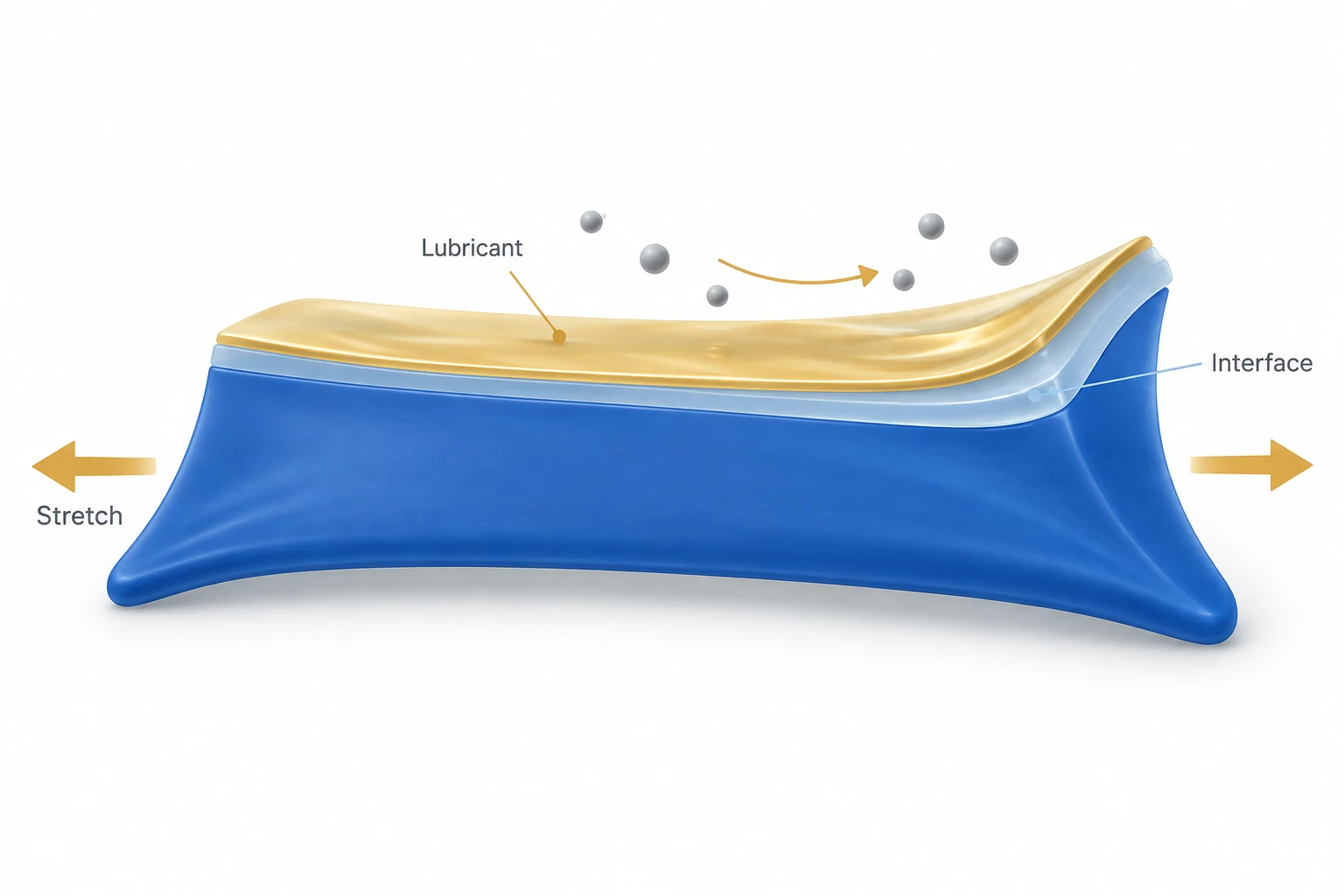
AI-generated concept: L-VIP aims to keep a lubricated interface continuous as the substrate stretches. Bands and arrows represent functions, not measured layers, thicknesses or particle paths. The image does not show experimental results or a maintenance-free implant lifetime.
Generated for this library to illustrate the design; it is not a figure from the paper and does not show measured data.
How it was evaluated
The coating was compared with a conventional non-stretchable lubricant-infused coating under 150% strain and applied to PVA hydrogel, filter paper, silicon wafer, polydimethylsiloxane (PDMS), Ecoflex, polyvinyl chloride, polypropylene and aluminum alloy. Wetting was measured with water, blood and other liquids, including during stretching to over 200% strain and after up to 2000 cycles at 150% strain. Mechanical and environmental durability were tested by a cross-cut adhesion test (ISO 2409) against PEG and polydopamine coatings, ultrasonication, water flushing, water shearing, toluene flow and repeated autoclaving, and dielectric performance by leakage current under accelerated aging and a simple motor demonstration. Fouling was assessed with albumin, fibrinogen, extracellular matrix molecules, Escherichia coli and Staphylococcus aureus, and cytocompatibility with NIH 3T3 fibroblasts. Coated and uncoated PVA hydrogels were implanted under the dorsal and hindlimb skin of 7-week-old female ICR mice for one and four weeks for histology, spleen and liver enzyme analysis, and retrieved samples were tested again after re-immersion in PFPE.
Key results
Under 150% strain, the conventional lubricant-infused coating cracked, whereas L-VIP remained crack-free. Coated materials showed similar static water contact angles regardless of the substrate, and coated filter paper repelled fetal bovine serum, dimethyl sulfoxide, blood, ethylene glycol and water. On Ecoflex stretched to 200% and tilted at 20°, blood droplets were pinned on the bare surface but left no residue on L-VIP. The coating maintained water contact angles of about 120° and sliding angles below 8° up to 200% strain, with partial defects and poorer wetting beyond that strain, and scanning electron microscopy showed no cracks after 2000 cycles at 150% strain, while the conventional coating cracked.
In the cross-cut test, the PEG coating peeled whereas none of the 25 VIP blocks detached. Fouling resistance to humic acid was nearly identical before and after 1 day of 20 kHz ultrasonication, seven days of water flushing at 5 mL/s and 30 days of water shearing at 1000 rpm, the coated microfluidic channel did not swell in toluene, and water contact angles changed insignificantly after nine autoclave cycles at 121°C. In the leakage current test, the coating without lubricant failed after 36 h, which the authors calculate as 20 days at 37°C, whereas L-VIP retained full dielectric performance over 1200 h, calculated as almost two years at body temperature; the abstract reports the accelerated aging test duration as over 50 days.
L-VIP reduced adhesion of albumin and fibrinogen compared with bare substrates and with a PEG coating, resisted fouling by extracellular matrix molecules including humic acid, and showed virtually no adherent E. coli or S. aureus after 24 h. Coated PVA hydrogels had no effect on fibroblast viability in indirect and direct culture and caused no significant change in fibroblast aspect ratio over five days.
In mice, neither implant produced abnormal skin reactions. After one week, tissue around bare PVA showed inflammation, a high density of macrophages and an organized thick collagen layer, whereas tissue around L-VIP-coated PVA showed a low inflammatory response, a significantly lower macrophage density and thin, low-density collagen; at four weeks the bare hydrogel was encapsulated by thick, dense collagen while the coated hydrogel was surrounded by few nuclei and little collagen. Spleen weight and length showed no significant change across groups at one and four weeks, and serum alanine and aspartate aminotransferase levels were similar with and without the coating after one week. Retrieved coated hydrogels separated from the subcutaneous tissue, and after re-immersion in PFPE they resisted albumin adhesion and showed no significant change in water contact angle compared with before implantation.
Open questions
Wetting deteriorated and partial defects appeared when strain exceeded 200%, which the authors attribute to the limited stretchability of the coating. The in vivo stability tests on retrieved hydrogels were performed after re-immersing them in PFPE. The authors acknowledge concerns about the bioaccumulation and environmental persistence of perfluorinated compounds; they estimate the perfluoropolymer film at about 100 nm thick and plan to explore alternative liquids for large-scale use.
Related research
Bioinspired self-repairing slippery surfaces with pressure-stable omniphobicity
SLIPS offers a comparison for how lubricant is retained; L-VIP asks how that interface holds up while a substrate stretches.
Abstract-based introductionRESEARCH NEWS
Research in the news
Unverified candidates and access limits (1)
Unverified candidate · [공과대학] 서정목 교수팀, 신축성과 유연성을 지닌 방오코팅기술 개발 ↗
Sources used in this summary
This summary is based on the full text of the published Wiley article, including the Experimental Section, Author Contributions and the legends of Figures 1-5; the Supporting Information was not reviewed. The accelerated aging condition is printed as 70° (no unit) and 10 V s−1 in the main text and as 10 V in the methods.