Overview
This study, by researchers at Brigham and Women’s Hospital, Harvard Medical School, and the BLISS Lab (Jungmok Seo) at Yonsei University, presents CARE, a customized wireless electrical stimulation patch for wounds that a 6-axis robot arm prints after a depth camera measures the wound. Pre-printing wound processing (scanning, wound extraction and size calculation) took 13.9 s to 17.0 s for 3 cm × 3 cm to 8 cm × 8 cm wounds, and simulations indicated a field of 88.8 mV/mm at 10 mm above the electrodes.
Background
Electrical stimulation can mimic or enhance the endogenous wound currents that guide cell migration and angiogenesis, but its clinical use has been limited by bulky hospital-based equipment, and patch-type devices must also adhere to tissue without damaging it on removal. Lithography and screen printing require masks and are poorly suited to wounds whose size and shape differ from patient to patient. Earlier in situ printing combined with 3D scanning used 3-axis systems that cannot easily reach wounds on the side of the body, and separate hand-held scanners need markers and coordinate transformations that add processing steps.
Design and approach
CARE is fabricated by INSIGHT, a system that mounts a depth camera and a pneumatic multi-syringe printhead on a 6-axis robot arm. The camera scans the body, the wound contour is extracted by color contrast with the surrounding skin, and a bounding box gives the wound width and length; curved or large wounds are captured from several angles and the point clouds are merged. The wound size is passed to a MATLAB script that sizes the interdigitated electrodes and the receiving coil so that the patch resonates with a 13.56 MHz excitation coil. Three inks, a silver conductive ink, a PDMS dielectric ink and a photocurable poly(glycerol-co-sebacate) acrylate (PGSA) adhesive, are then printed as five layers (coil, dielectric, electrodes, passivation and adhesive) on a flexible substrate on a hotplate at 80 °C, with the arm rotating to ±45° to switch between syringes.
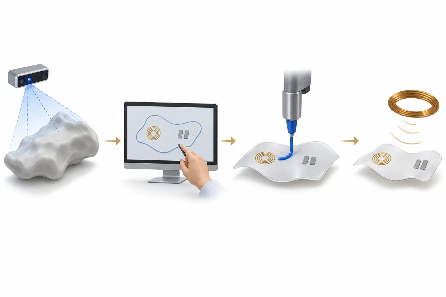
AI-generated concept, not to scale: CARE uses geometry information, a manually assisted design step and sequential printing with different inks. The scene shows one active nozzle at one point in the process. Electrodes, a receiving coil and a separate excitation coil symbolize functions; this is not a circuit diagram, experiment or treatment result.
Generated for this library to illustrate the design; it is not a figure from the paper and does not show measured data.
How it was evaluated
Scanning accuracy was tested on wound phantoms with five skin and five wound colors and on curved phantoms imaged from three angles (0° and ±30°). Printed conductive lines were cycled under 50% bending and 0–10% stretching, adhesion was measured by lap shear on porcine skin, and extract cytotoxicity was tested with human dermal fibroblasts (HDFs). Wireless performance was assessed by electric field simulation, inductance measurement and the transmission coefficient S21. Cellular effects of a 20 Hz pulsed radio frequency signal on a 13.56 MHz carrier were examined in HDFs and green fluorescent protein-expressing human umbilical vein endothelial cells (GFP-HUVECs) through proliferation, migration, tube formation, CD31 expression and growth factor secretion. Fit to an irregular wound was demonstrated ex vivo on the dorsal skin of a discarded mouse.
Key results
Pre-printing processing rose only from 13.9 s to 17.0 s as the wound size increased from 3 cm × 3 cm to 8 cm × 8 cm, and scanning finished within 20 s regardless of size. Width and length were recognized with less than 2.6% error across all 25 skin and wound color combinations, and multi-angle scans of curved phantoms gave 94% to 99% accuracy. Printing a patch took about 11 min for a 3 cm × 3 cm wound and approximately 42 min for an 8 cm × 8 cm wound, and the patch could be attached after 1 min of UV curing. Coils were also printed on curved substrates and on a substrate tilted at 45 degrees.
The resistance of printed conductive lines remained stable over 1000 bending cycles at 50% strain and increased gradually by up to 10% as tensile strain rose from 0 to 10%, returning to its original value on release. The PGSA adhesive gave a lap shear strength of 5.24 kPa on porcine skin, which the authors described as five times that of commercial fibrin glue (1.09 ± 0.04 kPa), and extracts at 100%, 50% and 25% concentration showed no significant difference in HDF viability.
Simulations indicated a field of 1.37 V/mm through the passivation layer and 88.8 mV/mm at 10 mm above the electrodes, above the 25 mV/mm level cited as promoting cell migration. The measured S21 was approximately 10 dB at 13.56 MHz, and about 3 V reached the patch when 10 V was applied to the external coil. For a 1.5 cm square wound, the target, optimized and measured coil inductances were 2.27, 2.03 and 1.75 μH; the authors attributed the lower measured value to removing half of the innermost turn to connect the electrodes.
In vitro, stimulation at a 99% duty cycle for 3 days yielded higher DNA content for both HDFs and GFP-HUVECs than controls. At a 20% duty cycle, migration was 1.86-fold faster for HDFs after 24 h and 1.64-fold faster for GFP-HUVECs after 8 h according to the Results, whereas the abstract gives factors of 1.6 for fibroblasts and 1.9 for endothelial cells. Tube formation after 6 h of stimulation produced over twice the number of junctions, CD31 expression increased 1.8-fold at a 99% duty cycle, and stimulated HDFs secreted more fibroblast growth factor (FGF) and vascular endothelial growth factor A (VEGF-A).
Open questions
The authors stated that the wound information was transferred manually to the MATLAB script and that a new algorithm is needed to design electrodes and coils automatically within the system. The excitation coil was on a rigid printed circuit board, and printing external coils on flexible substrates is required for translation. Future devices are planned to add temperature, impedance and pH sensors for closed-loop monitoring. Biological effects were evaluated in cell culture, with an ex vivo fit demonstration on a mouse.
Related research
Wireless, closed-loop, smart bandage with integrated sensors and stimulators for advanced wound care and accelerated healing
DOI: 10.1038/s41587-022-01528-3 ↗
The published abstract describes a wirelessly powered bandage that combines closed-loop monitoring of skin impedance and temperature with electrical stimulation and uses hydrogel electrodes that adhere and detach on demand, tested in mouse wound models. CARE addresses a different step: sizing and printing the electrodes and receiving coil to match each wound, with its biological effects examined in cell culture.
Abstract-based introductionRESEARCH NEWS
Research in the news
Sources used in this summary
This summary is based on the full text and figure legends of the NIH author manuscript in PMC (PMC11695167); the supporting information and movies were not reviewed, and the Methods describe the migration assay with different signal and timing values (about 10 Vpp at 4 Hz, 6 h) from those in the Results.