Overview
This study, developed by the BLISS Lab (Jungmok Seo) at Yonsei University with collaborators at Yonsei University and Hanyang University, introduces tannic acid (TA) into a poly(vinyl alcohol)/poly(acrylic acid) (PVA/PAA) double-network hydrogel to make a wound-dressing patch that is tough, self-healing, nonswellable, tissue-adhesive, hemostatic and antibacterial. The NHS-activated hydrogel showed a lap shear strength of about 35.98 kPa on porcine skin, and in a mouse liver puncture model the patch stopped bleeding within 10 s of application.
Background
Clips, staples and sutures remain the clinical standard for wound closure, but they do not reliably prevent leakage, bleeding or infection and can damage soft tissue. Bioadhesive glues such as fibrin glue and cyanoacrylate cure into brittle layers that fracture under cyclic tissue deformation, and adhesive films such as polyurethane are stiffer than soft tissue and detach during movement. Hydrogels match tissue softness, but they are usually brittle, adhere weakly, and swell in wet environments; double-network hydrogels are tougher, yet their dense polymer chains restrict self-healing, raise the modulus and increase water uptake. Commercial bioadhesives also provide only mechanical adhesion, and the authors note that reported hemostatic hydrogels require more than 90 s with steady pressure.
Design and approach
The hydrogel is made in two steps. TA, a polyphenol with 25 hydroxyl groups per molecule, is mixed into PVA to form a dynamic hydrogen-bonded network, and acrylic acid is then interpenetrated and polymerized with a disulfide crosslinker. The short PVA chains bound to TA dissipate energy, while the covalently crosslinked long PAA chains maintain integrity. Because both the TA hydrogen bonds and the disulfide bonds are reversible, the network can re-form after damage, and the hydrophobic moieties of TA shield the network from water. The dynamic network also makes the gel viscoelastic, so it can relax stress and fit rough tissue surfaces. For tissue bonding, the carboxyl groups of PAA are activated with 1-ethyl-3-(3-dimethylaminopropyl) carbodiimide and N-hydroxysuccinimide (EDC/NHS) to form covalent bonds with primary amines on tissue; a sodium bicarbonate and L-glutathione solution is used to release the patch. TA also contributes hemostatic and antibacterial activity.
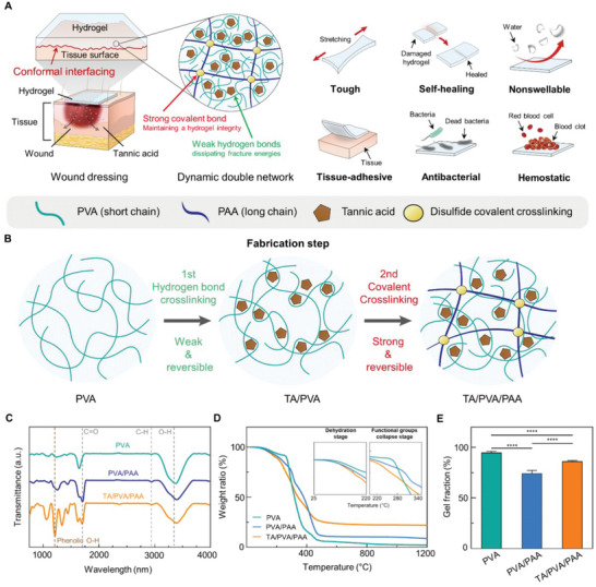
Double-network design, fabrication and material characterization of a TA/PVA/PAA wound-dressing hydrogel.
Jae Park, Tae Young Kim, Yeonju Kim et al. “A Mechanically Resilient and Tissue‐Conformable Hydrogel with Hemostatic and Antibacterial Capabilities for Wound Care”. Figure 1. DOI: 10.1002/advs.202303651. CC BY 4.0. Figure content is unchanged.
Source figure and caption ↗ · DOI: 10.1002/advs.202303651 ↗ · CC BY 4.0 ↗
How it was evaluated
The TA/PVA/PAA hydrogel was characterized by X-ray photoelectron spectroscopy, Fourier transform infrared spectroscopy, thermogravimetry, gel fraction, water retention, tensile, cyclic, notch and self-healing tests, swelling in water, rheology and stress relaxation at 15% strain, with single-network PVA and conventional PVA/PAA double-network hydrogels as comparisons in the spectroscopic, thermogravimetric, gel fraction, tensile, swelling and stress relaxation tests. Adhesion was measured by lap shear on porcine skin. Cytocompatibility was tested with NIH 3T3 fibroblasts in a transwell system, hemolysis with horse erythrocytes, and the blood clotting index (BCI) with whole blood from ICR mice. Antibacterial activity was assessed against Escherichia coli and Staphylococcus aureus by inhibition zone tests and live/dead staining. In seven-week-old female ICR mice, patches were implanted subcutaneously for 7 days for histology and liver enzyme measurement, and applied to livers punctured with an 18-gauge needle, with untreated livers and a PVA patch as comparisons and blood collected on filter papers at 10 and 120 s.
Key results
The gel fraction of TA/PVA/PAA was about 86.05%, compared with about 74.39% for PVA/PAA and about 94.85 for PVA, and the equilibrium water content was about 45.29% and was retained over 24 h. With 20% w/v TA, which the authors selected for its high stretchability and sufficient tensile strength (raising TA up to 30% w/v increased tensile strength but reduced strain at break, and higher concentrations reduced tensile strength), the hydrogel stretched to about 1109% strain with a tensile strength of about 119 kPa, whereas PVA reached about 108 kPa but less than about 310% strain. The Young's modulus was 18.65 ± 3.78 kPa, and cyclic loading showed similar energy dissipation over five cycles.
Notched samples elongated beyond 750% strain without crack propagation. Cut pieces recovered about 40% of their tensile strength after 30 s and 100% after 300 s, while PVA, PVA/PAA and alginate/poly(acrylamide) hydrogels without TA did not self-heal. The TA/PVA/PAA hydrogel retained its volume after 24 h in water with no mass increase, whereas the swelling ratio reached about 500% for PVA/PAA and about 177% for PVA. Under a constant 15% strain, TA/PVA/PAA relaxed to half its initial stress within a few seconds, while PVA and PVA/PAA showed no decrease.
On porcine skin, the NHS-activated hydrogel reached a lap shear strength of about 35.98 kPa, against about 8.28 kPa for the non-activated hydrogel and about 12.72 kPa after the trigger solution; the abstract reports this adhesion as about 31 kPa. The bonded hydrogel elongated by about 200% during shear without cohesive failure, compared with about 61% after the trigger solution, and stayed attached when the skin was stretched, twisted and bent.
In vitro, the hydrogels showed no significant toxicity toward fibroblasts at 1, 3 and 5 days with or without NHS, and the hemolysis ratio of about 11% did not differ significantly from the phosphate-buffered saline control. The BCI of TA/PVA/PAA was 58.40 ± 1.5, approximately two times lower than the control, PVA and PVA/PAA groups. TA-containing hydrogels produced inhibition zones of about 8.03 ± 0.7 and 9.83 ± 2.59 mm against E. coli and 11.8 ± 0.62 and 11.5 ± 1.47 mm against S. aureus, whereas PVA and PVA/PAA produced none. In mice, subcutaneous patches caused no significant change in macrophage density or serum alanine and aspartate aminotransferase levels after 7 days. In the liver puncture model, the TA/PVA/PAA patch stayed in place under bleeding pressure, reduced blood mass significantly compared with untreated and PVA groups, and induced hemostasis within 10 s, with dense red blood cells, platelets and fibrin threads on its surface.
Related research
Tough adhesives for diverse wet surfaces
DOI: 10.1126/science.aah6362 ↗
Li and colleagues' abstract distinguishes bonding at the tissue surface from energy dissipation inside an adhesive. The TA-containing patch adds hemostatic and antibacterial functions to its network.
Abstract-based introductionRESEARCH NEWS
Research in the news
Unverified candidates and access limits (1)
Unverified candidate · 어디에든 붙이면 지혈·자가치유 가능한 패치 개발 / YTN 사이언스 ↗
Sources used in this summary
This summary is based on the open-access full text in Europe PMC (PMC10602564), including the Experimental Section and the legends of Figures 1-6; the Supporting Information was not reviewed. The original article gives the inhibition zone incubation as 24 h in the results text and 12 h in the Figure 5 legend and methods.