Overview
This review, written by the BLISS Lab (Jungmok Seo) at Yonsei University, examines bioadhesive hydrogels as interfaces that regulate immune and regenerative responses in chronic wounds rather than as simple coverings. It links mechanisms such as macrophage polarization, NF-κB, NLRP3 and STAT signaling and interfacial mechanotransduction to four engineering strategies, and notes, for example, that tabulated hemostatic bioadhesives span wet adhesion strengths of 5.29 to 35.98 kPa.
Background
Chronic wounds, common in aging and diabetic patients, remain in a prolonged inflammatory state in which macrophages fail to shift from the M1 to the M2 phenotype. The review describes this block as the convergence of signals from neutrophil extracellular traps, advanced glycation end-products, dendritic cells, Th17 cells and senescent fibroblasts on the macrophage, sustained by exudate, biofilm, hypoxia, abnormal pH and protease activity. Gauze, films, foams, hydrocolloids, alginates and conventional hydrogel dressings each serve one or a few passive functions, and many lift or detach from wet, irregular and moving wound surfaces.
How the review is organized
The review defines immunomodulation broadly, including indirect effects such as antioxidant and antibacterial activity as well as direct regulation of macrophage polarization and cytokines. It sets out required properties for clinical use: wet adhesion and conformal contact, toughness and fatigue resistance, a soft hydrated structure, biocompatibility and immune tolerance, controlled permeability and moisture handling, and stimuli responsiveness. It argues that these should be prioritized by wound type, such as diabetic foot ulcers, venous leg ulcers, pressure injuries and ischemic wounds, and matched to each healing stage. Candidate signaling routes linking hydrogel properties to healing include NF-κB, STAT3/STAT6, Nrf2/HO-1, integrin–FAK, YAP/TAZ and Piezo1.
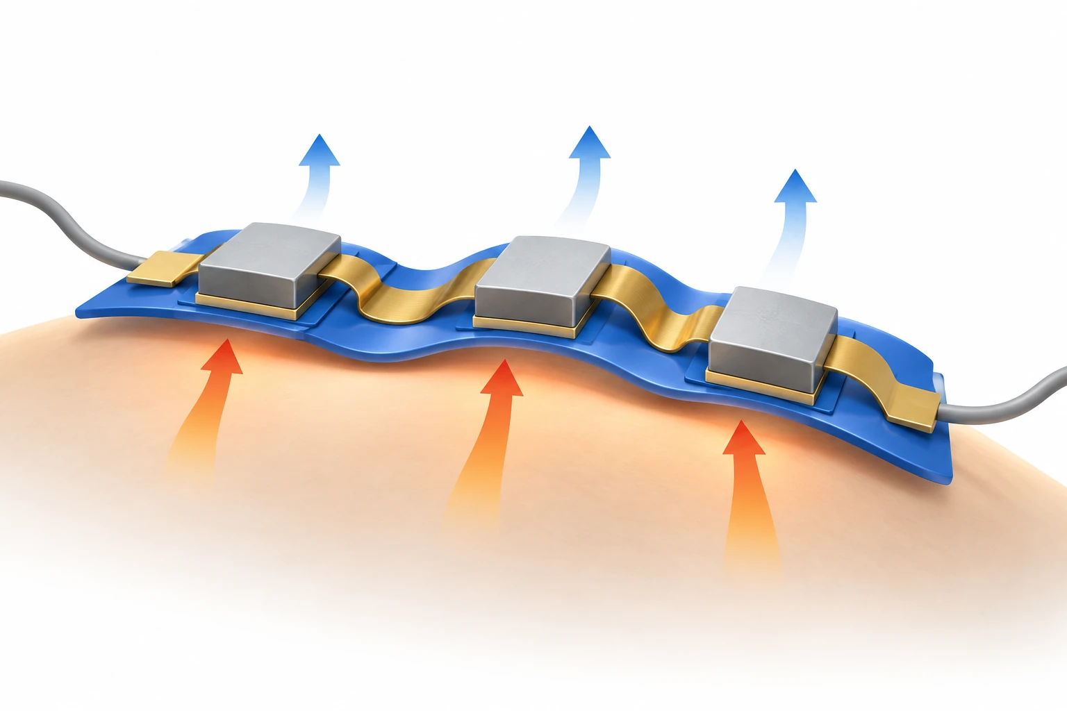
AI-generated concept: the review connects hydrogel adhesion and removal, deformation, transport and the timing of immune regulation at chronic wounds. Shapes and proportions are illustrative, not experimental evidence of material performance or clinical efficacy.
Generated for this library to illustrate the design; it is not a figure from the paper and does not show measured data.
Scope of the review
The review organizes the literature into structural and protective hydrogels (shielding and sealing, exudate control, hemostasis, cooling), bioactive and regenerative hydrogels (microbe- and cell-laden systems, controlled delivery of drugs, growth factors, exosomes and metal ions), external-stimuli-based hydrogels (electrical stimulation, ultrasound and near-infrared or optical phototherapy) and sensing-integrated hydrogels (temperature, pH and impedance). Table 1 compares six conventional dressing types with bioadhesive immunomodulatory hydrogels. Table 2 lists hemostatic bioadhesives by gelation time, wet adhesion strength, immunomodulatory outcome and animal models; Table 3 lists stimulus-responsive hydrogels by stimulus condition and wound model; and Table 4 compares sensing platforms by sensing mechanism, operational stability and performance under exudate and biofouling.
Main points of the review
For hemostatic bioadhesives, the review reports wet adhesion strengths of 5.29 to 35.98 kPa and gelation times from 1.7 s to patches pre-cured for 1 h, and argues that the appropriate kinetics depend on the delivery route. It cautions against direct numerical comparison across animal models: the same 1.7 s gelation of one adhesive gave hemostasis in 59 s in rats but about 2.1 min in pigs. It also notes that highly exuding chronic wounds produce 0.5 to 1.0 ml of exudate per 10 cm2 per day and that an optimal swelling range has not been defined.
Among stimulus-based systems, the review describes conductive hydrogels for electrical stimulation, including a spray-printed liquid-metal patch in which 2 mA direct current for 4 h per day reduced the M1 marker CD86 while preserving CD206, as well as piezoelectric hydrogels that generate reactive oxygen species under ultrasound and light-activated hydrogels. It notes that electrical platforms were mostly tested in uninfected models, whereas ultrasound and phototherapy systems were tested in infected burn or diabetic models, so outcomes should be compared within wound type.
For sensing-integrated dressings, the review notes that infected wounds tend toward pH above 7.5 and that temperature and impedance track inflammation and barrier recovery. It finds that seven-day measurements of drift, sensitivity retention and biofouling are rarely reported and that most in vivo studies show correlations rather than calibration against reference methods.
On translation, the review identifies reliance on acute rodent models as a major limitation and proposes stepwise validation through disease-relevant and splinted rodent models, porcine models and ex vivo human skin. It notes that hydrogel dressings without an added drug or biologic may fall within the US Class I, 510(k)-exempt category, whereas antimicrobial, biologic, cell-containing or electronic products face more demanding pathways, and it expects near-term translation to favor simple, scalable formulations with durable wet adhesion and a limited number of mechanistically validated functions.
Open questions
The review states that the relative contribution of each signaling pathway should be interpreted with caution and calls for pathway-specific inhibitors, knockdown or reporter systems to establish causal links. It lists unresolved issues including adhesion durability under wet and dynamic conditions, the trade-off between leak-proof sealing and atraumatic removal, long-term safety of electronic and living components, sterilization, reproducibility, manufacturing cost and regulatory classification.
Related research
Wireless, closed-loop, smart bandage with integrated sensors and stimulators for advanced wound care and accelerated healing
DOI: 10.1038/s41587-022-01528-3 ↗
A mouse system combining sensing, stimulation and detachable hydrogel provides an example of the review's adaptive-design approach.
Abstract-based introductionRESEARCH NEWS
Research in the news
Sources used in this summary
This summary is based on the full text and Table 1 of the open-access review (PMC13602266); Tables 2 to 4 were reviewed through their headings and the passages that cite them, and the cited primary studies were not read.