Overview
The STAT (Strong Tissue-adhesive And Thrombostatic) hydrogel, developed by the BLISS Lab (Jungmok Seo) at Yonsei University, is a double-network hydrogel in which partially oxidized catechol groups bond to wet tissue without a separate activation step, and tannic acid promotes clotting. The paper reports adhesion "within seconds without any pretreatment", a lap-shear strength of 273 kPa on porcine skin, and hemostasis within 10 s in a mouse liver puncture model, with bleeding also controlled in rabbit spleen and liver injuries.
Background
Clips, staples and sutures take time to apply, depend on operator skill and often fail to seal high-flow bleeding, particularly in deep or irregular wounds. Patch-type hydrogel dressings cover large areas but conform poorly to complex cavities, whereas liquid glues reach confined sites but can be washed out before a clot forms. Shear-thinning double-network (DN) hydrogels combine toughness with injectability, but they generally adhere weakly to tissue; covalent bonding is usually added through carbodiimide (EDC/NHS) activation, which takes more than 1 h and leaves potentially cytotoxic byproducts.
Design and approach
The first network consists of poly(vinyl alcohol) (PVA), dopamine-functionalized sodium alginate (SA-DOPA), tannic acid (TA) and borax, which provide reversible hydrogen bonds and borate ester links for toughness and self-healing. Poly(acrylic acid) (PAA) is then polymerized within this network as a ductile second network. Gentle kneading in air partially oxidizes the SA-DOPA catechols to o-quinone; borax suppresses premature oxidation during static storage, and oxidation proceeds only under mixing. The quinones bond covalently to tissue amines and thiols through Michael-type addition, while TA interacts with platelets and fibrin to accelerate clotting and contributes antibacterial activity. The same material can be used as a preformed patch or extruded through a syringe.
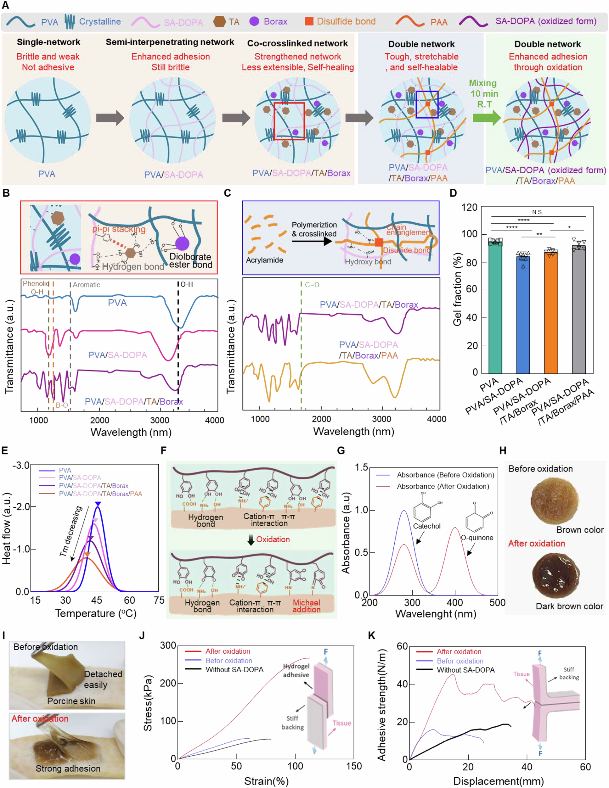
Network formation, oxidation, structural characterization and porcine-skin adhesion measurements for STAT.
Tae Young Kim, Kayoung Son, Chang-Hwan Moon et al. “Quinone-mediated, tissue-adaptive double-network hydrogel for instant hemostasis and wet-tissue adhesion”. Figure 2. DOI: 10.1038/s41467-026-72068-6. CC BY 4.0. Figure content is unchanged.
Source figure and caption ↗ · DOI: 10.1038/s41467-026-72068-6 ↗ · CC BY 4.0 ↗
How it was evaluated
Adhesion was measured on porcine skin by lap shear (ASTM F2255) and 180° peel (ASTM F2256) tests, with amine blocking by sulfo-NHS acetate to test the bonding mechanism, and by rheometry as a function of mixing time. Mechanical tests covered tensile, cyclic, stress-relaxation, notch and self-healing behavior. In vitro work included inhibition-zone assays against E. coli and S. aureus, NIH3T3 fibroblast viability, hemolysis and blood clotting index. Sealing was demonstrated in an ex vivo porcine gastric perforation under endoscopic guidance. In vivo, the patch was tested in a mouse liver puncture model (18G needle) against untreated and gauze groups, and in rabbit spleen incision and liver dissection models against gauze, fibrin glue (Tisseel) and TachoSil. The injectable form was compared with sutures, fibrin glue and a zip-type closure band in a 2.0 cm mouse dorsal skin incision over 14 days.
Key results
Oxidation of catechol to o-quinone increased adhesion to porcine skin by more than two-fold in both lap shear and 180° peel tests; in the representative lap-shear curves the peak stress rose from 52.9 kPa (catechol state) to 267.6 kPa (o-quinone state), and Supplementary Table 1 lists a shear adhesion strength of 273 kPa. Blocking tissue amines reduced the adhesion of oxidized STAT from 260–284 kPa to 9–13 kPa, consistent with quinone–amine coupling. Adhesion increased with kneading time up to 15 min and declined at 20 min, which the authors attributed to water loss. On timing, the paper states that STAT achieves robust adhesion within seconds; the 10 s value given in the main text and in Supplementary Table 1 refers to hemostatic time.
STAT showed a tensile strength of almost 150 kPa, an elongation of about 580% and a Young's modulus of about 80 kPa, within the range of soft tissues. Notched specimens still stretched beyond 600% with a notch of 20% of the width, and cut specimens recovered more than 90% of their original strength after 10 s of contact. The hydrogel sealed a 1 × 1 cm² perforation in an ex vivo porcine stomach after endoscopic delivery.
Only TA-containing hydrogels produced inhibition zones against E. coli and S. aureus. STAT reached a blood clotting index of 58.40 ± 1.7, about half that of TA-free controls, and was non-hemolytic and non-cytotoxic to NIH3T3 fibroblasts. In the mouse liver puncture model, hemostatic time was 1–3 s with STAT versus 95–110 s with gauze, and blood loss was 8–28 mg versus 206–240 mg in the untreated and gauze groups (n = 3). In rabbits, gauze, fibrin glue and TachoSil left active bleeding after 3 min of compression, whereas STAT stopped bleeding within seconds; blood loss with STAT was 1.35–2.21 g (liver) and 3.8–4.3 g (spleen), compared with 6.9–8.2 g and 14.9–17.2 g for gauze.
In the mouse skin incision model, the syringe-applied hydrogel kept the wound nearly closed by day 7 and fully recovered by day 14, with aligned epidermis, continuous collagen and few inflammatory cells on histology. The tensile strength of healed skin at day 14 was 889–942 kPa with STAT and 409–438 kPa with sutures (P = 0.0059); both remained below uninjured skin (1154–1276 kPa).
Open questions
The authors report that mixing beyond 15 min reduced both adhesion and mechanical properties because of water loss, and that oxidation continues during storage in air, so samples were stored under N₂ to preserve adhesion for one week. Healed skin at day 14 remained weaker than native dermis owing to ongoing remodeling.
Related research
Dry double-sided tape for adhesion of wet tissues and devices
DOI: 10.1038/s41586-019-1710-5 ↗
The publisher abstract describes a dry tape that absorbs interfacial water before forming covalent bonds. It offers a mechanistic comparison with hydrated STAT.
Abstract-based introductionRESEARCH NEWS
Research in the news
서정목 교수팀, 젖은 조직에 즉시 접착·지혈하는 이중네트워크 하이드로겔 개발 ↗
연세대, 젖은 조직에도 신속 접착·지혈 소재 개발 ↗
연세대 서정목 교수팀, 젖은 조직에 즉시 접착·지혈하는 하이드로겔 개발 ↗
연세대학교 서정목 교수팀, 10초 만에 젖은 조직 붙이고 출혈 막는 차세대 하이드로겔 개발 ↗
STAT Hydrogel Stops Bleeding in Seconds Without Sutures ↗
Jungmok Seo: A Quinone-Activated Hydrogel for Second-Scale Hemostasis and Suture-Free Surgical Sealing ↗
[연구 프론티어] 서정목 교수팀, 젖은 조직에 즉시 접착·지혈하는 이중네트워크 하이드로겔 개발 ↗
Sources used in this summary
This summary is based on the full text and figure legends of the open-access article (PMC13287715), the Supplementary Information figure legends and Supplementary Table 1, and selected Source Data sheets; values given as ranges are the individual data points in the Source Data. The supplementary movies, reporting summary and peer review file were not reviewed.