Overview
This study, developed by the BLISS Lab (Jungmok Seo) at Yonsei University with collaborators, applied a coating termed TAB (targeting-specific interaction and blocking nonspecific adhesion) to thermally drawn multifunctional neural fibers. The coating combines a perfluoropolyether lubricant layer with immobilized brain-derived neurotrophic factor (BDNF), reduced the maximum insertion load into a brain phantom from 25.2 to 2.94 mN, and supported single-unit recording in the mouse hippocampus for up to 12 months, whereas bare fibers no longer recorded spikes after 6 months.
Background
Implanted neural probes lose recording quality over time because of insertion trauma, chronic foreign body reaction (FBR), glial encapsulation and micromotion at the device-tissue interface. Antifouling coatings reduce protein and immune cell adhesion, but strong repellency also prevents contact with the neurons that must be recorded. Conversely, surfaces conjugated with neurotrophic biomolecules promote neuronal interaction, yet the biomolecules are exposed to proteases and activated immune cells, which degrade them, and such coatings do not address frictional damage during insertion.
Design and approach
The fiber is thermally drawn from a polycarbonate/poly(methyl methacrylate) (PC/PMMA) preform together with tungsten microwires, giving a 276 ± 1.52 μm fiber that contains an optical waveguide, a microfluidic channel and recording electrodes. The TAB coating is formed by partial silanization: the surface is first fluorinated with perfluorosilane (PFS), a brief secondary O2 plasma (30 W, 5 s) removes part of the fluorinated layer, and the exposed sites are functionalized with 3-aminopropyltriethoxysilane (APTES). BDNF is then covalently coupled to the amine groups by EDC/NHS chemistry, and a PFPE lubricant is infused. The lubricant is retained on the PFS regions, while BDNF on the APTES regions remains accessible to TrkB receptors on neurons and astrocytes.
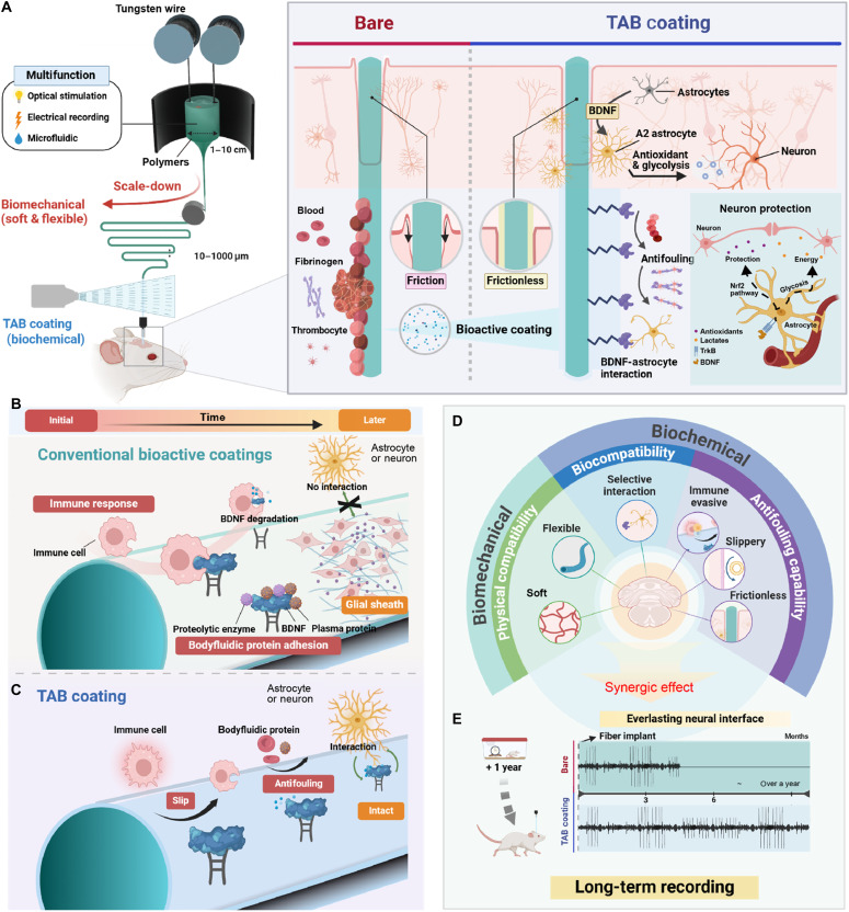
Flexible neural-fiber construction, TAB versus conventional coatings, and mechanical/biochemical functions.
Tae Young Kim, Yeonzu Son, Keun-Young Yook et al. “Bioadaptive liquid-infused multifunctional fibers for long-term neural recording via BDNF stabilization and enhanced neural interaction”. Figure 1. DOI: 10.1126/sciadv.adz1228. CC BY-NC 4.0. Figure content is unchanged.
Source figure and caption ↗ · DOI: 10.1126/sciadv.adz1228 ↗ · CC BY-NC 4.0 ↗
Noncommercial academic use requires author/source attribution, the license link and disclosure of changes.
How it was evaluated
The coating was characterized by XPS, contact angle, antifouling tests with horse blood and fluorescent plasma proteins (fibrinogen, albumin), insertion and retraction loads in a 0.6% agarose brain phantom, and a cross-cut tape test (ASTM D3359). Molecular dynamics and density functional theory calculations examined lubricant distribution on PFS/APTES surfaces. Primary mouse hippocampal cells were cultured on bare, lubricant-only, BDNF-only and TAB surfaces to assess cell selectivity, cytokine and MMP-9 release, and antioxidant and glycolysis gene expression. In mice, FBR was examined after subcutaneous implantation (n = 3 per group, 7 days), immunohistochemistry was performed at 1 day to 12 weeks after hippocampal implantation, and single-unit activity in CA1 was recorded for up to 12 months.
Key results
In the brain phantom, the TAB fiber reduced the maximum insertion load from 25.2 to 2.94 mN and the relaxed load after insertion from 3.94 to 1.45 mN relative to the bare fiber, and also lowered the retraction load. The coating remained intact after 10 tape-peeling cycles, whereas a poly(ethylene glycol)-based antifouling coating delaminated completely after 5 cycles. The coated fiber stayed free of blood staining and showed no plasma protein adhesion in fluorescence images. Molecular dynamics simulations indicated 7.6-fold higher lubricant localization on the fluorinated regions than on the APTES regions, consistent with BDNF exposure between lubricated domains.
In primary hippocampal cultures, astrocytes and neurons adhered and spread on both BDNF-only and TAB surfaces, but microglia on TAB surfaces remained rounded and showed lower signal intensity, whereas microglia on BDNF-only surfaces became ramified. MMP-9, TNF-α, IL-6 and IL-8 were elevated only in the BDNF-only group without lubricant. qPCR and Western blot analyses showed increased expression of genes and proteins associated with the TrkB/Nrf2 antioxidant pathway and with glycolysis on the TAB surface.
After implantation in the mouse hippocampus, MMP-9 expression was elevated around BDNF-only fibers on day 1, and Iba1-positive activated microglia were reduced around TAB fibers at week 1. The neuroprotective A2 astrocyte marker S100A10 was higher at the TAB interface at day 1 and week 1, while the A1 marker C3 was not elevated. The NeuN-positive neuronal area around TAB fibers was larger than around bare fibers at 1, 4 and 12 weeks, with no difference on day 1. In the subcutaneous model, TAB fibers showed less immune cell infiltration and collagen deposition after 1 week.
TAB fibers recorded single-unit activity in hippocampal CA1 for up to 12 months, whereas waveform, principal component and interspike interval analyses indicated that bare fibers were no longer recording spikes after 6 months. Over the statistically analyzable 8-month period, the number of spike units per electrode decreased for bare fibers (P = 0.016, n = 6 electrodes) and was maintained for TAB fibers (P = 0.796, n = 5 electrodes), and the SNR of TAB recordings did not change significantly (P = 0.447, n = 3).
Related research
Multifunctional fibers for simultaneous optical, electrical and chemical interrogation of neural circuits in vivo
The publisher abstract and metadata describe Canales and colleagues' drawn fibers for recording, optical functions and fluid delivery. TAB focuses additionally on surface biochemistry and host response.
Abstract-based introductionRESEARCH NEWS
Research in the news
서정목 교수팀, 뇌-컴퓨터 인터페이스 장기 안정성 극복할 신경 전극 코팅 기술 개발 ↗
Bioadaptive Liquid-Infused Multifunctional Fibers for Long-Term Neural Recording via BDNF Stabilization and Enhanced Neural Interaction ↗
뇌·컴퓨터 인터페이스 신호 1년 이상 기록한다 ↗
[22일 대학가] 세종대·중앙대·한양대·연세대 ↗
New paper on neural recording technology published in Science Advances ↗
Device–tissue interfaces: prior studies, ELFS biliary stents and FLUID ureteral stents ↗
Unverified candidates and access limits (1)
Sources used in this summary
This summary is based on the full text, figure legends and Materials and Methods of the open-access article (PMC12422202); the supplementary materials were not reviewed. Quantitative values for in vitro cell coverage and protein attachment are taken from the Introduction, and most histological comparisons are reported as relative differences without numerical values in the main text.