Overview
This in vitro study, by researchers at the BLISS Lab (Jungmok Seo) at Yonsei University and UNIST with collaborators at Incheon National University, introduced the precision drug testing organ chip (PreD chip), in which perfluoropolyether (PFPE) lubricant coats the polydimethylsiloxane (PDMS) channel walls while cells grow on a separate extracellular matrix (ECM)-coated membrane. In cell-free channels, the conventional PDMS chip lowered the outflow concentration of dexamethasone by about 30%, whereas the PreD chip showed no noticeable loss, and a gut model in the PreD chip gave a dexamethasone IC50 of about 350 ± 19.7 μg mL−1 compared with 400 ± 23.8 μg mL−1 in the conventional chip.
Background
Organ-on-a-chip (organ chip) devices reproduce cell–cell and tissue–tissue interfaces under flow and are being developed as human-relevant preclinical models. PDMS is the most widely used material for these devices, but its porosity allows absorption of small molecules of 100 to 1000 Da, and more than 60% of commercially available drugs are hydrophobic small molecules, so drug exposure in the chip can differ from the dosed concentration. Thermoplastic chips resist absorption but are rigid and difficult to bond into complex structures, and additive or surface modifications of PDMS can alter its mechanics or lose hydrophilicity over time. A PFPE lubricant coating had prevented absorption in PDMS devices, but filling the whole channel with coating solution also coated the cell culture membrane.
Design and approach
The PreD chip stacks upper and lower PDMS channels with a porous polyethylene terephthalate (PET) membrane (2.0 μm pores). After oxygen plasma treatment, the PDMS parts are silanized with 3-aminopropyltriethoxysilane (APTES) and coated with a carboxyl-terminated perfluoropolymer (PFP), while the PET membrane is treated with 3-glycidoxypropyltrimethoxysilane (GPTES). Thermal curing at 170 °C forms amide bonds between APTES and PFP and β-hydroxyamine bonds between APTES and the GPTES-treated membrane, and the PFP layer is then swollen with PFPE lubricant (Krytox GPL 101). The membrane is coated with ECM proteins, so that the lubricated channel walls repel drugs and proteins while the cells attach to the membrane.
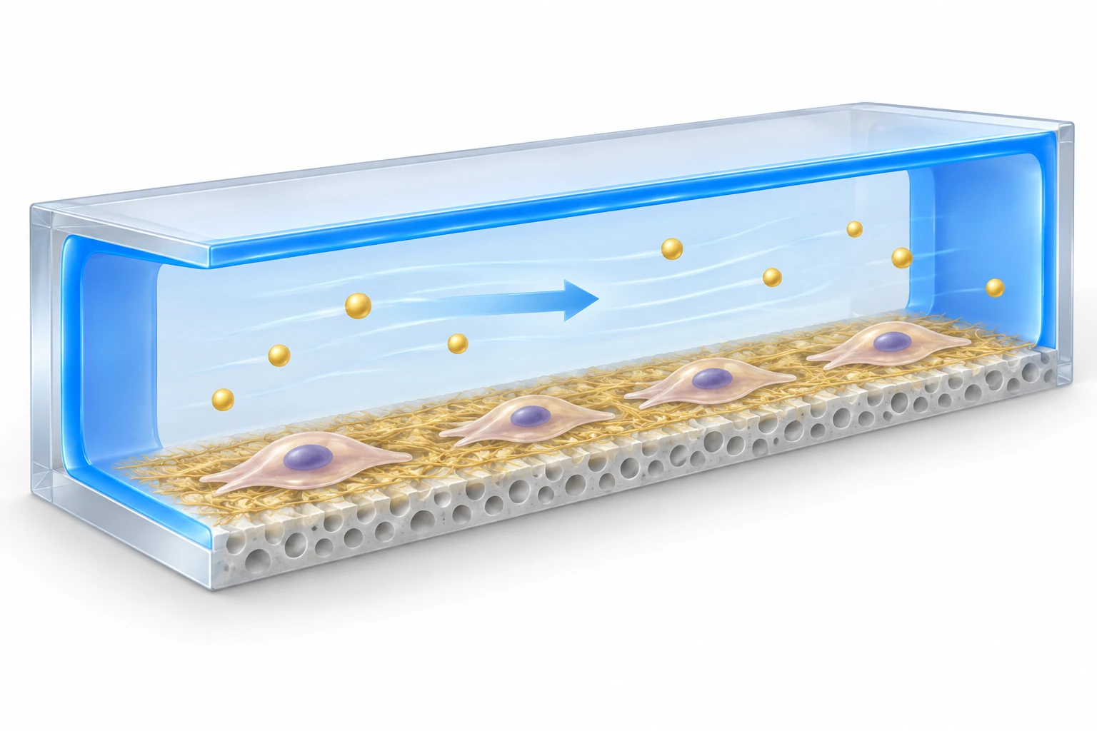
AI-generated concept: PreD's lubricated channel walls and ECM-coated PET membrane serve different roles. Only one simplified channel is shown. The image does not present drug-recovery measurements, toxicity predictions or organ-function results.
Generated for this library to illustrate the design; it is not a figure from the paper and does not show measured data.
How it was evaluated
Absorption was tested with rhodamine B (479 Da) and with dexamethasone (DEX, 392 Da) at 400 to 1000 μg mL−1 in cell-free channels at 30 μL hr−1, with inflow and outflow measured by high-performance liquid chromatography. The coating was characterized by X-ray photoelectron spectroscopy, bonding tensile tests, contact angles with water, fetal bovine serum, blood, ethylene glycol and dimethyl sulfoxide, blood and plasma protein fouling, toluene exposure, lubricant retention under PBS flow of 50 to 200 μL hr−1, and O2 and CO2 permeability of a 50 μm PDMS film. A gut model was formed with Caco-2 cells and used for DEX toxicity tests at 100 to 500 μg mL−1 for one day, and a blood–brain barrier (BBB) model was formed with human induced pluripotent stem cell-derived brain endothelial cells (iPS-BECs), primary human pericytes and astrocytes and used for dopamine (153 Da) and levodopa (197 Da) permeability tests, each compared with a conventional PDMS chip.
Key results
Rhodamine B diffused into the conventional PDMS channel over time, whereas the PreD chip resisted absorption after 1 day of flow. In cell-free channels, the PreD chip showed no noticeable DEX loss at 400 to 1000 μg mL−1, while the conventional chip reduced the outflow concentration by about 30% at all tested concentrations. The lubricated PDMS showed similar contact angles for liquids with different surface tensions, let blood droplets slide without residue, showed significantly low albumin and fibrinogen adsorption and did not swell in toluene. The water contact angle remained similar after 15 days in PBS at 37 °C, more than 80% of the lubricant remained after 9 days of PBS flow, and 8 to 9 of every 10 chips were fabricated successfully.
The PFP coating reduced O2 and CO2 permeability of the PDMS film to about 70%, which improved slightly after lubricant swelling; the text also describes the reduction with lubricant swelling as about 60%. Caco-2 cells cultured for 7 days at 30 μL hr−1 showed no significant change in the hypoxia-related genes HIF1A, VEGF, SOX2 and CD42 compared with the conventional chip. The gut epithelium formed villi-like structures expressing chromogranin A and villin, with villi heights of 123.7 ± 11.7 μm in the PreD chip and 127.3 ± 18.7 μm in the conventional chip, similar CCK-8 viability, and transepithelial electrical resistance (TEER) that increased over 7 days as in the conventional chip.
After one day of DEX exposure, the IC50 was about 350 ± 19.7 μg mL−1 in the PreD chip and 400 ± 23.8 μg mL−1 in the conventional chip, which the authors describe as an increase in toxicity sensitivity of about 12.5%; a static Transwell gut model did not reach the IC50 even at 500 μg mL−1. At 300 μg mL−1, villi height decreased slightly in the PreD chip without statistical significance, and at 400 μg mL−1 villi height was 48 ± 11.3 μm in the PreD chip compared with 78.3 ± 9.3 μm in the conventional chip.
In the BBB model, barrier function and cell viability did not differ significantly from the conventional chip, and iPS-BECs in both chips formed tight junctions containing ZO-1 and Claudin-5 and expressed Occludin and GLUT-1. In empty chips, the PreD chip recovered about 94.9% of dopamine and 98.6% of levodopa, compared with 83.2% and 89.6% in the conventional chip (n = 4). Dopamine permeability across the BBB was similar in both chips, whereas levodopa permeability was significantly higher in the PreD chip, so the difference between the BBB-impermeable and BBB-permeable drugs was clearer.
Open questions
The authors state that the clinical relevance of the PreD chip requires comprehensive validation with a wider range of drugs, including responses and transport across tissue barriers, and that cell types with high oxygen demand, such as hepatocytes, may need further optimization of the lubricant coating and perfusion conditions.
Related research
PDMS absorption of small molecules and consequences in microfluidic applications
Toepke and Beebe describe changes in concentration as small molecules partition into PDMS. PreD responds through channel coating and separate cell-culture regions.
Abstract-based introductionRESEARCH NEWS
Research in the news
Sources used in this summary
This summary is based on the published open-access PDF of the article, including the main text, figure legends and Experimental Section; the Supporting Information was not reviewed.