Overview
This study, co-authored by Jungmok Seo (then at the School of Electrical and Electronic Engineering, Yonsei University), who now leads the BLISS Lab at Yonsei University, moved individual water droplets on a thin superhydrophobic polydimethylsiloxane (PDMS) micro-pillar film by pulling a local dimple into it with vacuum from below. Spreading the pillars lowered the water adhesion force from 67 to 49 μN, and droplets followed the moving dimple along programmed paths for transport, merging and mixing.
Background
Continuous-flow microfluidic devices need pumps, valves and mechanical mixers and suffer from unintended flow patterns, limited flow velocities and a lack of reconfigurability. Droplet-based alternatives driven by electrowetting, light, magnetic fields, electrostatic forces or dielectrophoresis require additives such as magnetizable particles or external field sources, and droplet handling on superhydrophobic surfaces had been limited to a small set of simple operations.
Design and approach
A 275 μm thick PDMS film with round-tipped micro-pillars (2.5 μm radius, 4 μm height, 4.5 μm period) was cast in one moulding step from a silicon mould coated with dodecyltrichlorosilane and was superhydrophobic without further treatment, with a water contact angle (WCA) of 151 ± 3°. The film was suspended over a holder, and a vacuum tip beneath it, moved by a programmed translational stage, pulled down a hemispherical dimple. Stretching and curvature spread the pillars at the positive curvature of the dimple border and packed them at the negative curvature of the bottom. Because the adhesion force (Fadh) on such a Cassie-Baxter surface scales with the number of pillars in contact, the border has lower local adhesion, and once the slope-induced gravitational force exceeds it, the droplet detaches there and follows the moving dimple.
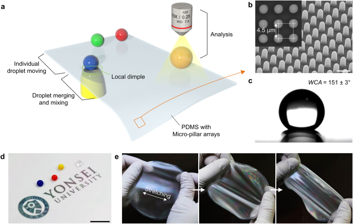
The figure presents the micropillared PDMS platform, its local dimples for moving, merging and analysing droplets, and substrate characterization.
Jungmok Seo, Seoung-Ki Lee, Jaehong Lee et al. “Path-programmable water droplet manipulations on an adhesion controlled superhydrophobic surface”. Figure 1. DOI: 10.1038/srep12326. CC BY 4.0. Figure content is unchanged.
Source figure and caption ↗ · DOI: 10.1038/srep12326 ↗ · CC BY 4.0 ↗
How it was evaluated
Dimple shapes were examined with SU-8 negative replicas and PDMS duplicates imaged by SEM, at a fixed pressure difference of −81 kPa and vacuum tip diameters from 2 to 3.75 mm. Fadh was measured with a home-made microelectromechanical balance using a 10 μl droplet on PDMS substrates fabricated with pillar spacing strains (εdist) of 0, 25, 50 and 75%. The platform was applied to surface-enhanced Raman spectroscopy (SERS) of rhodamine 6G (R6G) from 10^-3 to 10^-15 M with about 80 nm Ag nanoparticles, and to formulating lipidoid (ND98)-siRNA complexes against green fluorescent protein (GFP) for transfection of GFP-expressing HeLa cells, compared with complexes mixed by manual pipetting (flow cytometry, n = 3).
Key results
With a 2.85 mm tip, the substrate stretched by 20.7%, and the pillar spacing, 2 μm in the flat region, changed by up to 49% at the positive curvature and −11% at the negative curvature. Increasing the tip diameter from 2 to 3.75 mm raised stretching from about 7 to about 32%, reduced the absolute positive bending radius from about 0.8 to 0.4 mm and increased the negative bending radius from about 1.3 to 1.8 mm, so that εdist could be tuned from −15 to 61%; measured values agreed with calculations.
Fadh decreased from 67 to 49 μN as εdist increased up to 50%, which the authors attribute to fewer pillars contacting the droplet. A 10 μl droplet followed the dimple along an N-shaped path, droplets were merged and fully mixed by moving them back and forth, and larger tips could move smaller droplets. The pillar density differed by more than 50% between the edge and centre of the dimple, and the minimum droplet volume that could be controlled was about 7 μl.
In SERS, droplets of R6G and Ag nanoparticles merged on the surface gave a detection limit of 10^-5 M in situ, limited by freely diffusing molecules and particles. Evaporating the merged droplet on the superhydrophobic surface concentrated the analyte and allowed ex situ detection down to 10^-15 M, which the authors report as a hundred-fold lower limit than conventional SERS on a flat substrate. Droplet mixtures could be removed without residue, allowing repeated measurements on the same substrate.
Two days after transfection of GFP-HeLa cells, complexes mixed by pipetting silenced GFP by 70.0 ± 1.9%, whereas complexes formed by merging and mixing droplets on the surface for 5 min silenced GFP by 78.8 ± 0.5% (n = 3, p < 0.01); the abstract reports this as a transfection efficiency of about 80%.
Open questions
The authors note that the ex situ SERS detection limit was poorer than earlier reports on superhydrophobic nanosensors because the PDMS micro-pillar arrays retain some water adhesion, and suggest that a more water-repellent surface could concentrate analytes into a smaller area.
Related research
Bioinspired self-repairing slippery surfaces with pressure-stable omniphobicity
Slippery liquid-infused porous surfaces (SLIPS) lock an infused lubricating fluid into nano/microstructured substrates to repel simple and complex liquids with low contact angle hysteresis, whereas the present platform keeps droplets on a dry superhydrophobic micro-pillar surface and moves them by deforming the substrate locally.
Abstract-based introductionRESEARCH NEWS
Research in the news
Sources used in this summary
This summary is based on the open-access full text and figure legends (PMC4511949); the Supplementary Information and Supplementary Movie 1 were not reviewed.