Overview
This is a study by researchers at Sungkyunkwan University, the Institute for Basic Science and the BLISS Lab (Jungmok Seo) at Yonsei University on a resealable antithrombotic artificial vascular graft (RAAVG) made from a tough self-healing polymer (T-SHP) conduit with a lubricious inner coating and an integrated carbon nanotube (CNT) pressure sensor. The graft resealed after 25G needle puncture under blood circulation, a sutured interface reinforced with a second T-SHP layer withstood 50% strain without cracks, and the self-bonded sensor followed flow-induced pressure changes at flow rates of 10 to 100 mL/min.
Background
Coronary artery bypass grafting relies on autologous vessels, which may be unavailable in infants and the elderly, and commercially available expanded polytetrafluoroethylene (ePTFE) grafts lack a lubricious lumen and are unsuitable for small vessels because of thrombosis, blockage and infection. The authors also note that conventional grafts lack self-healing and self-sealing after mechanical damage such as needling, that covalently cross-linked elastomers such as PDMS cannot dissipate the stress at tightly sutured interfaces, and that blood pressure sensors attached by gluing or suturing suffer from mechanical mismatch and detachment.
Design and approach
The T-SHP is a PDMS-based polymer cross-linked by strong dynamic hydrogen bonds of 4,4′-methylenebis(phenylurea) (MPU) units and weak hydrogen bonds of isophorone bisurea (IU) units. A drop-cast T-SHP film is rolled around a mold and heals into a seamless conduit at 60 °C for 1 h, and its lumen is coated with polydopamine (PDA), a perfluoropolymer (PFP) cured at 80 °C for 3 h, and a perfluoropolyether (PFPE) lubricant that swells the PFP to form an inner layer of about 200 nm mimicking the tunica intima. The MPU:IU ratio of 2:8 (M0.2I0.8) was chosen because the 4:6 film was too stiff to wrap around small bending radii and the 0:10 film showed irreversible viscous stretching. The pressure sensor (P-CNT) is a single-walled CNT network embedded in T-SHP, so it attaches to the conduit by film-to-film self-healing.
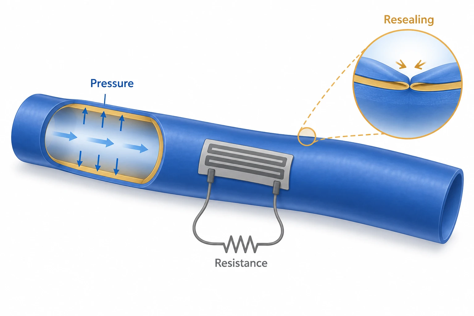
AI-generated concept separating resistance sensing of conduit pressure or deformation from resealing after damage.
Generated for this library to illustrate the design; it is not a figure from the paper and does not show measured data.
How it was evaluated
T-SHP films with MPU:IU ratios of 4:6, 2:8 and 0:10 were tested in tension and for self-healing in PBS. RAAVG surfaces were compared with PTFE, ePTFE and uncoated T-SHP for contact and sliding angles of water, artificial cerebrospinal fluid, simulated body fluid and blood, and tested for blood cell adhesion (including comparison with a heparin coating), albumin and fibrinogen adsorption, lubricant retention under shaking at 100, 200 and 300 rpm, degradation in PBS and simulated body fluid at 37 °C over 10 days, and fibroblast cytotoxicity by extract assay. Citrated horse blood was circulated for 2 h through bare and coated silicone tubing (36 mL/min in the Results; 37 mL/min in the Methods) to measure thrombus weight and residual platelets. Hemostasis was tested by 25G needle puncture of a RAAVG in a closed blood circulation loop, and suture tests used PDMS or T-SHP films sewn to porcine aorta. The P-CNT sensor was characterized for gauge factor, response to 10, 50 and 100 kPa, flow-induced resistance changes and repeated needle puncture. Feasibility was examined ex vivo by suturing RAAVG to porcine aorta and in rats by subcutaneous implantation for one week and by wrapping RAAVG around a punched inferior vena cava of a male Sprague-Dawley rat for 3 days, each compared with a sham group.
Key results
The Young's moduli of the 4:6, 2:8 and 0:10 films were 0.54, 0.41 and 0.33 MPa and their toughness values were 18.95, 5.49 and 1.97 J m−2; the authors describe all moduli as comparable with those of rodent, primate and human vessels, citing 0.2−0.6 MPa for human vessels in the physiological range. T-SHP films healed in PBS for 4 h stretched beyond 200% strain without fracture propagation, and RAAVG conduits kept their shape after 30 pinching cycles.
Fluid droplets were trapped on PTFE and ePTFE and blood did not slide on them, whereas RAAVG films showed sliding angles below 10° and kept their sliding angles after shaking at up to 300 rpm. Blood cells and clots adhered to T-SHP but not to RAAVG, and RAAVG showed almost no adhesion where a heparin-coated substrate showed a small amount. Albumin and fibrinogen adsorption on RAAVG was negligible after 2 h of incubation. In the blood circulation test, the lubricant-coated tubing yielded less thrombus and more residual platelets, and the introduction reports that only 70% of platelets remained with bare tubing. Neither T-SHP nor RAAVG degraded over 10 days, extract media did not reduce fibroblast viability, and a RAAVG cut in half and healed still repelled blood.
In the closed circulation loop at 50 mL/min, removing a 25G needle left a single blood droplet with no further leakage, and the puncture site disappeared after 24 h of circulation at room temperature. A sutured single-layer T-SHP film deformed without fracture at 30% strain, whereas a PDMS film (1:25 precursor to cross-linker) broke; with a second T-SHP layer healed over the suture, the interface showed no cracks or holes at 50% strain.
The less conductive P-CNT sensor had gauge factors of 0.184 and 0.186 below and above 30% strain, against 0.053 and 0.140 for the more conductive sensor, and it distinguished pressures of 10, 50 and 100 kPa. Mounted on a T-SHP conduit, it gave average resistance amplitudes of 3.93, 9.57 and 13.17 kΩ at maximum flow rates of 10, 50 and 100 mL/min according to the Results text (the Fig. 5 legend lists 20, 50 and 100 mL/min). Leakage stopped immediately after needle withdrawal, sensing at 100 mL/min remained stable through five needle punctures without electrical disturbance, and the sensor followed randomly changing flow rates of 20, 50 and 100 mL/min. Razor-blade scratches on the sensor healed, and integrating the sensor slightly increased the Young's modulus relative to bare T-SHP.
Ex vivo, horse blood flowed through porcine aorta sutured to RAAVG without leakage. After one week of subcutaneous implantation in rats, hematoxylin and eosin (H&E) staining showed no biodegradation and no significant inflammatory response compared with controls. After 3 days wrapped around a punched inferior vena cava, RAAVG covered the hole without significant inflammation compared with the sham group, and SEM showed no significant adhesion on its inner and outer surfaces.
Open questions
Because graft surgery is difficult in a small animal model, the in vivo vascular test wrapped RAAVG around a punched inferior vena cava instead of replacing a vessel segment, and it lasted 3 days. The authors also note that the self-healing of the graft material can glue the conduit walls together upon pinching, which the lubricious inner coating prevents.
Related research
Biodegradable and flexible arterial-pulse sensor for the wireless monitoring of blood flow
DOI: 10.1038/s41551-018-0336-5 ↗
Boutry and colleagues describe a biodegradable, flexible capacitive sensor wrapped around an artery that reports the arterial pulse wirelessly. This study combines a resistive CNT pressure sensor with the graft itself, using the same self-healing polymer so that the sensor bonds to the conduit and both reseal after needle puncture.
Abstract-based introductionRESEARCH NEWS
Research in the news
Sources used in this summary
This summary is based on the published ACS Nano PDF, including the abstract, Introduction, Results and Discussion, Conclusion, Experimental Section and the legends of Figs. 1-6; the Supporting Information (Figs. S1-S10 and Movies S1-S4) was not reviewed.