Overview
This study reports a 3D-printable hydrogel ink with incorporated nanomaterials (THIN) whose stiffness and conductivity are set by the choice of nanofiller, developed by the BLISS Lab (Jungmok Seo) at Yonsei University. A Laponite XLG nanoclay version (THIN-X) serves as a soft tissue-interfacing layer and a functionalized carbon nanotube version (THIN-C) as the conductor, giving a Young's modulus range of 10–300 kPa and a conductivity of about 20 S/m, and the two inks were printed into a ring-shaped strain sensor worn on the fingers.
Background
Soft electronics need conformal contact with tissue for accurate biosignal monitoring, but elastomers such as PDMS (0.5–3 MPa) and polyurethane (6 MPa) are much stiffer than tissue (20–100 kPa) and, being dry and non-viscoelastic, adhere poorly to soft substrates. Soft hydrogels match tissue better but are brittle and lack conductivity. Nanofillers can reinforce hydrogels and add conductivity, yet they tend to restrict the chain mobility needed for self-healing, swelling degrades electrical and adhesive performance, and fabrication methods for precise hydrogel structures remain limited.
Design and approach
THIN combines a double network of poly(vinyl alcohol) (PVA) and poly(acrylic acid) (PAA) with tannic acid (TA). PVA provides short, energy-dissipating chains, PAA forms covalently crosslinked long chains with disulfide crosslinks, and TA acts as a dynamic crosslinker through reversible hydrogen bonds that supports flowability, viscoelasticity and self-healing. THIN-X (PVA:TA:XLG = 10:10:1) uses XLG nanoplatelets to raise the modulus and retain shape and serves as a tissue-interfacing layer that keeps current in the conductor layer from flowing directly into the body, while THIN-C (PVA:TA:fCNT = 40:40:1) uses functionalized carbon nanotubes (fCNTs) to form a conductive pathway. N-hydroxysuccinimide (NHS) ester groups on PAA bond covalently to amine groups on tissue, and the shear-thinning inks are printed at 95 °C and joined through self-healing.
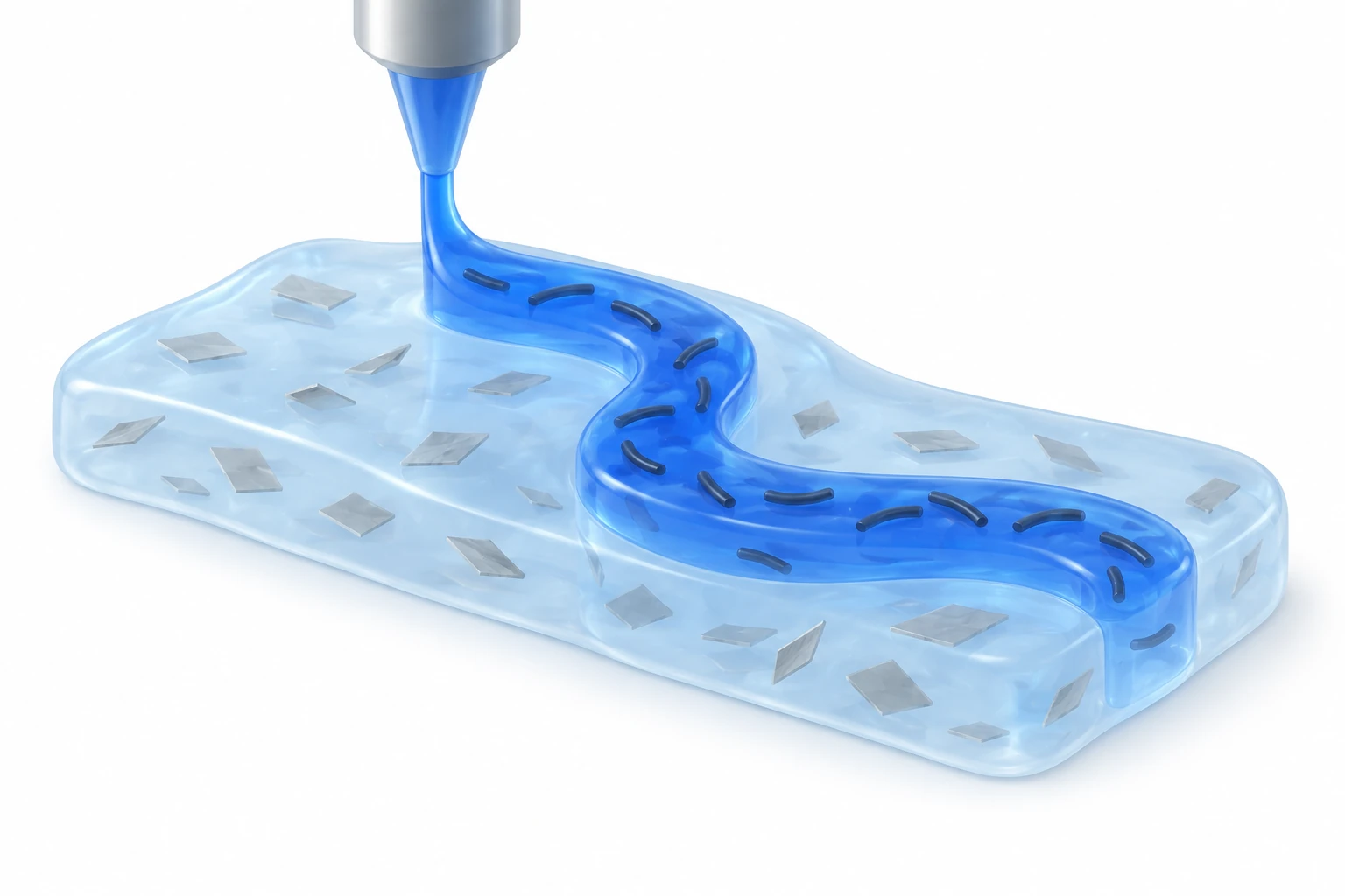
AI-generated concept: a nanoclay-containing THIN-X support is distinguished from a functionalized-CNT THIN-C conductive path. The image explains material roles rather than reproducing a formulation, filler distribution, device geometry or recovery result.
Generated for this library to illustrate the design; it is not a figure from the paper and does not show measured data.
How it was evaluated
The inks were characterized by tensile tests with notches of 20% and 40% of the width, self-healing tests in air and in water, swelling in water for up to 24 h and in phosphate-buffered saline (PBS) for 14 days, cyclic loading at 10%, 30% and 50% strain, and rheology over 25 to 95 °C. Biocompatibility was assessed with NIH-3T3 fibroblasts in transwell culture and with Caco-2 cells for 5 days. Adhesion was measured by lap shear on porcine skin and on glass, PDMS, titanium (Ti) and Pi substrates. THIN-C was tested for conductivity, impedance between platinum electrodes and resistance under stretching, and a printed THIN-X/THIN-C ring sensor was worn on the finger joints to drive a virtual hand.
Key results
Cut THIN-X recovered 30% of its mechanical properties after 10 s and returned to its original state after 5 min; hydrogels re-attached in deionized water for 5 min separated away from the healed site when pulled. A 20% notch caused no significant difference in strain-at-break. Without TA, PVA showed a 200% increase in swelling ratio after 24 h, whereas THIN remained non-swellable even after 14 days in PBS. The energy release rate was 53.7 J/m2 for THIN-X and 39.3 J/m2 for THIN-C.
Both inks were shear-thinning, and the storage modulus exceeded the loss modulus throughout the temperature scan, so the printed structures retained their shape; a resolution of up to 100 μm was achieved and printed THIN-C retained a water content of 91%. Fibroblast viability with THIN-X or THIN-C showed no significant difference from the control without hydrogel after 5 days.
NHS-functionalized THIN-X reached a lap shear strength of about 22 kPa on porcine skin, compared with 4.91 kPa without NHS and 6.55 kPa after treatment with a sodium bicarbonate and L-glutathione trigger solution used for detachment. Bonding strength did not change significantly between 5 min and 48 h, and values of 13 to 36 kPa were obtained on glass, PDMS, Ti and Pi. Under a constant 15% strain, THIN-X and THIN-C relaxed to half their initial stress within a few seconds, and THIN-X conformed to a curved surface after 10 min.
THIN-C reached about 20 S/m at the percolation threshold, maintained its conductivity in PBS for 14 days, and showed an impedance of about 200 Ω between 10^2 and 10^5 Hz. Its gauge factor was about 1.12 for strains up to 80%, within the 0.46 to 1.51 range of previous hydrogel strain sensors cited by the authors. The text states both that resistance returned to its initial value after 5 s and that the recovery time was 2 s. In the ring sensor, printed with 2,160 μm (THIN-X) and 1,800 μm (THIN-C) nozzles, each finger's sensor responded when that finger was bent, and the signals moved the corresponding fingers of a virtual hand in a hand simulator game.
Open questions
The authors reported that applying 400% strain decreased the conductivity of THIN-C by about 3 S/m, suggesting irreversible cleavage of the conducting pathway, and that the resistance of printed THIN-C increased as the nozzle diameter decreased, so the printed line thickness had to be chosen for conductivity.
Related research
3D printing of conducting polymers
DOI: 10.1038/s41467-020-15316-7 ↗
Yuk and colleagues print intrinsically conductive PEDOT:PSS structures and neural electrodes. THIN instead adjusts a CNT-filled hydrogel network.
Description drawing on the main textSources used in this summary
This summary is based on the main text, figure legends and Materials and methods of the published version of record (Biosensors and Bioelectronics, 13-page Elsevier PDF, local copy). The supplementary data (Figures S1–S28, Table S1 and Movies S1–S4) were not reviewed; values attributed to supplementary figures are those stated in the main text.