Overview
This study, developed by the BLISS Lab (Jungmok Seo) at Yonsei University with the Department of Biotechnology at Yonsei University, presents a 3D-printable hydrogel ink of poly(vinyl alcohol) (PVA), tannic acid (TA) and poly(acrylic acid) (PAA) in which a double network of weak and strong hydrogen bonds provides shear-thinning extrusion, toughness and self-healing. The optimized ink was printed at a resolution of about 100 μm and showed a tensile strength of 45.6 kPa and an elongation at break of 656%, and with carbon nanotube (CNT) fillers it reached a conductivity of about 0.95 S/m.
Background
Hydrogels reduce the mechanical mismatch between electronics and tissue, but they are often weak and brittle under large deformation and lack self-healing. Earlier self-healing, printable conductive hydrogels, such as a polyaniline/poly(4-styrenesulfonate) network and a conductive adhesive cellulose ink, were printed at low resolution (over 0.6 mm) and not extended to 3D structures, and a tough printable agar/polyacrylamide double-network hydrogel lacked self-healing and high printing resolution.
Design and approach
PVA provides a soft, biocompatible base; TA forms weak, reversible hydrogen bonds with PVA, and PAA, cross-linked with the disulfide-containing N,N′-bis(acryloyl)cystamine, forms strong hydrogen bonds as a second network. During printing the ink is heated to 85 °C so that weak bonds break and the ink flows, and the bonds reform immediately after deposition so that the printed structure keeps its shape. Carboxyl-functionalized CNTs are dispersed into the PVA/TA solution before the PAA network is formed, and for tissue adhesion the hydrogel is functionalized with N-hydroxysuccinimide (NHS) ester groups.
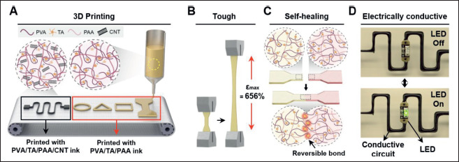
3D printing, stretching, self-healing and conductive-circuit demonstrations of multimaterial hydrogel inks.
Soo A Kim, Yeontaek Lee, Kijun Park et al. “3D printing of mechanically tough and self-healing hydrogels with carbon nanotube fillers”. Figure 1. DOI: 10.18063/ijb.765. CC BY 4.0. Figure content is unchanged.
Source figure and caption ↗ · DOI: 10.18063/ijb.765 ↗ · CC BY 4.0 ↗
How it was evaluated
Inks with PVA:TA mass ratios of 1:0.5, 1:1 and 1:2 were compared by shear-rate and temperature (25 to 95 °C) rheology, with PVA/PAA without TA included in the temperature sweep, and by swelling in deionized water over 960 min, printing fidelity with a 600 μm nozzle and tensile tests of printed samples; the 1:1 ink was further printed with 400, 200 and 100 μm nozzles and compared with bulk hydrogel in tensile tests. Self-healing was quantified from tensile strength after contact times of 30 to 300 s. CNT concentrations of 1, 3, 6 and 9 mg/mL were tested for mechanical properties and for conductivity by a modified four-point probe. Adhesion to porcine skin and the viability of NIH-3T3 fibroblasts over 5 days (n = 3) were examined, and printed circuits were used for LED lighting, resistance recovery after cutting, gauge factor and responses to compression, bending and stretching.
Key results
At 85 °C all inks showed shear-thinning. PVA/PAA without TA remained elastic (G′ larger than G″) across the tested temperatures and its moduli dropped sharply near 80 °C, whereas PVA/TA/PAA inks had close G′ and G″ values that decreased continuously with temperature. The 1:1 ink had the highest viscosity, about 100 Pa·s at 100 s−1, and moduli of about 10 kPa at 85 °C, and it showed almost 0% swelling over 960 min, whereas the 1:0.5 hydrogel swelled to 7.5% within 240 min and the 1:2 hydrogel first decreased to −3.7% within 100 min before expanding. Water contents of printed 1:0.5, 1:1 and 1:2 hydrogels were about 70.8%, 92.4% and 83.5%.
The 1:1 ink formed uniform filaments of nozzle-like width through a 600 μm nozzle, 2D shapes through nozzles down to 100 μm, meshes with a 100 μm nozzle and self-supporting multilayer structures, all without post-processing. Its maximum tensile strength and elongation at break were 45.6 kPa and 656% (about 650% in the abstract). Printed samples reached an elongation of up to 600% with tensile strength similar to the bulk hydrogel, but a lower Young's modulus than the bulk, which the authors attribute to dehydration after printing; it remained tissue-like at about 15 kPa. Separated samples rejoined into a stretchable structure after 3 min of contact, with a healing efficiency of 30% after 30 s and 80% after 5 min, and in tests after 180 s of healing the hydrogel elongated to 890% at 85 °C.
Increasing the CNT concentration raised the tensile strength and reduced the elongation at break. At 6 mg/mL the conductivity was about 0.95 S/m, above the 0.3–0.7 S/m range the authors cite for human tissue, and this concentration was used for further tests. NHS-treated hydrogels adhered to porcine skin after pressing for 5 min, and fibroblast viability with the hydrogel did not differ significantly from controls over 5 days.
A printed CNT hydrogel circuit lit an LED that turned off when the circuit was cut and turned on again when the cut ends were rejoined, and the resistance returned to its initial value after self-healing. The gauge factor was 1.356 for 0–300% strain and 4.457 for 300–400% strain, and resistance change ratios reached about 30%, 10% and 100% under compression, tensile bending and stretching.
Open questions
The authors report trade-offs within the tested formulations: printed hydrogels had a lower Young's modulus than bulk hydrogels, attributed to dehydration after printing, and adding CNTs reduced the elongation at break compared with the hydrogel without CNTs.
Related research
3D printing of conducting polymers
DOI: 10.1038/s41467-020-15316-7 ↗
Yuk and colleagues print PEDOT:PSS conductive paths together with insulating materials. This hydrogel study prints a PVA/TA/PAA network with CNT fillers and examines how conduction returns when a cut printed path is rejoined by self-healing.
Description drawing on the main textSources used in this summary
This summary is based on the open-access full text in PubMed Central (PMC10406165), including the Materials and methods, Results and discussion and the legends of Figs. 1-5; the supplementary file (Figs. S1-S11 and Table S1) was not reviewed.