Overview
This review, co-authored by Jungmok Seo (then at the Nanobio Device Laboratory, School of Electrical and Electronic Engineering, Yonsei University, with a current address at Brigham and Women's Hospital, Harvard Medical School and the Harvard-MIT Division of Health Sciences and Technology), who now leads the BLISS Lab at Yonsei University, surveys how natural surfaces with extreme wettability are reproduced in engineered materials and how these surfaces are used in biomedicine. It links surface chemistry and micro/nanostructure to applications in cell patterning, three-dimensional (3D) spheroid culture, antifouling medical devices, open-channel droplet microfluidics and high-throughput cell assays.
Background
Superhydrophilic and superhydrophobic surfaces have attracted attention for potential uses in self-cleaning fabrics, anti-fog windows, anti-corrosive coatings, drag reduction and water transport. Controlling wettability through surface chemistry and structure can also create cellular microenvironments and cell–substrate interactions that conventional culture platforms do not provide, but the authors note that only a few reports had examined the roles and applications of these surfaces in the biomedical field.
How the review is organized
The review first sets out the wetting principles, Young's equation and the Wenzel and Cassie-Baxter models, and defines superhydrophobic surfaces as those with a water contact angle (CA) above 150°. It then pairs each natural model with fabrication strategies: the hierarchical, wax-covered lotus leaf for water repellence, the rose petal for water adhesion, the rice leaf and Morpho butterfly wing for anisotropic and unidirectional wetting, the Stenocara desert beetle for patterned wettability and water collection, and the Nepenthes pitcher plant for lubricant-infused slippery surfaces, followed by surfaces whose adhesion switches with temperature, light, curvature or gas.
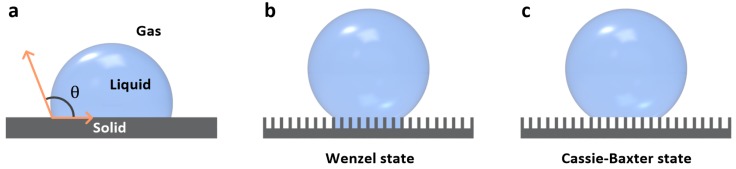
The review schematic compares droplet contact on a flat surface and in the Wenzel and Cassie–Baxter wetting states.
Sera Shin, Jungmok Seo, Heetak Han et al. “Bio-Inspired Extreme Wetting Surfaces for Biomedical Applications”. Figure 2. DOI: 10.3390/ma9020116. CC BY 4.0. Figure content is unchanged.
Source figure and caption ↗ · DOI: 10.3390/ma9020116 ↗ · CC BY 4.0 ↗
Scope of the review
Section 3 organizes biomedical applications into five areas: cell patterning for studies of cell–cell and cell–material interactions, hanging-drop culture of functional cell spheroids, antibacterial and anti-thrombogenic surfaces for medical devices, open-channel droplet-based lab-on-a-chip systems, and high-throughput cell and biomaterial assays. For each area, the review summarizes representative studies with their cell types, test conditions and reported outcomes, and closes with challenges and future directions.
Main points of the review
Among fabrication examples, the review describes vertically aligned silicon nanowire (Si NW) surfaces whose water CA changed from 0 to 154° after rapid thermal annealing at 1000 °C, and Pd-coated Si NW arrays whose droplet adhesion switched between air and H2 atmospheres. It notes that UV-driven adhesion switching is less suitable for biological use because UV irradiation affects enzymes, cells and chemicals.
For cell culture, the summarized studies include hydrophilic patterns on superhydrophobic substrates where cells in neighbouring spots communicated directly below a spacing of 250 μm, a screen of 24 alginate-based hydrogel compositions, and polydopamine (pDA)-patterned hanging drops in which mesenchymal stem cell spheroids secreted about 300% more vascular endothelial growth factor (VEGF) than spinner flask cultures and rat islet cell spheroids showed about 200% higher glucose sensitivity. On Pd-coated Si NW arrays, spheroid size of human adipose-derived stem cells was set by cell density (1.25, 2.5 and 5.0 × 10^5 cells/mL) and medium volume (10, 15 and 20 μL), with viability maintained after four days.
For medical devices, the cited results include superhydrophobic xerogel coatings that reduced S. aureus and P. aeruginosa adhesion by 99% and 98%, structured polymer surfaces that after 24 h with E. coli gave fewer than 100 colony-forming units (CFUs) on polystyrene (PS) and polyethylene (PE) and none on polycarbonate (PC), compared with 100,100 CFUs on flat PS and PC and 25,800 on flat PE, slippery surfaces that prevented biofilm accumulation for over one week and reduced bacterial attachment by 96%–96.6% relative to polyethylene glycol surfaces, and tethered-liquid perfluorocarbon (TLP) coatings that reduced occlusive thrombosis in tubing after 8 h of flow in vivo.
For droplet microfluidics and screening, the review reports a pDA-patterned slippery device that handled organic solvents with surface tension above that of the lubricant (17.1 mN/m) and raised a reaction yield to 70.3% from 52.1% in a 4 mL vial, a vacuum-actuated superhydrophobic PDMS platform that detected R6G at 10−5 M in droplets and 10−15 M after evaporation and gave a transfection efficiency of 78.8% ± 0.5% compared with 70.0% ± 1.9% for manual mixing, arrays of up to 85,000 hydrogels formed in several minutes, and superhydrophobic scaffolds with 36 biomaterial combinations implanted subcutaneously in Wistar rats.
Open questions
The authors state that biomedical uses of bio-inspired wetting surfaces remain at an early stage. Surface durability and long-term stability must be improved, because continuous contact with culture medium causes protein adsorption and liquid penetration into the micro/nanostructures that change wetting properties. They point to self-healing and stimuli-responsive composites, surfaces that capture and release biochemicals, and scalable fabrication by spray coating, stamping and 3D printing as directions for further work.
Related research
Bioinspired self-repairing slippery surfaces with pressure-stable omniphobicity
Slippery liquid-infused porous surfaces (SLIPS) provide a retained-lubricant example for comparing the wetting designs discussed in the review.
Abstract-based introductionRESEARCH NEWS
Research in the news
Sources used in this summary
This summary is based on the open-access full text in Europe PMC (PMC5456462), including figure captions, checked against the publisher PDF; the individual studies cited in the review were not read.