Overview
This study, developed by the BLISS Lab (Jungmok Seo) at Yonsei University with collaborators, describes FLUID, a ureteral stent in which a silicone primer and silicone oil are integrated into a fully microporous thermoplastic polyurethane (TPU) wall that acts as a lubricant reservoir. In an artificial ureter, FLUID showed a peak pull-out force of 0.07 ± 0.02 N without pre-hydration, and in a porcine model it reduced magnesium and calcium deposition by 84.1% and 45.1% at 4 weeks relative to a commercial stent.
Background
Long-term ureteral stents are limited by friction against the urothelium, protein conditioning films, bacterial colonization and mineral encrustation, and clinical indwelling is generally limited to 3 to 6 months. Commercial stents use softening materials, smooth surfaces or hydrophilic coatings, yet encrustation persists. Hydrated polymer and hydrogel coatings can swell, delaminate or lose function under abrasion, and conventional slippery liquid-infused porous surfaces (SLIPS) hold lubricant only in thin surface microstructures that are difficult to form on long, narrow tubular devices and are depleted under shear.
Design and approach
The stent core is extruded from TPU with a foaming agent, which produces micropores throughout the wall (average total porosity 6.24%). A silicone elastomer primer is dip-coated under vacuum so that it covers the outer surface and the internal pore walls, and medical-grade silicone oil (350 cSt, selected from 50 to 10,000 cSt by friction tests) is then dip-coated onto the primer. The primer anchors the oil through chemical affinity, and oil held in the pore network redistributes to the surface after local depletion. The mobile oil layer is intended to limit direct solid contact and the stable attachment of proteins, bacteria and mineral nuclei.
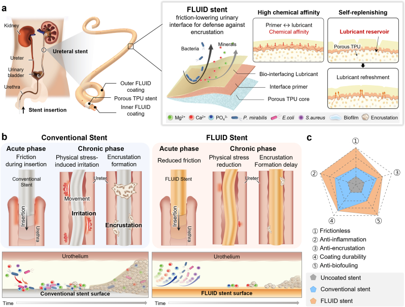
FLUID ureteral-stent lubricant storage and design comparisons during insertion and indwelling.
Yejin Jo, Yeontaek Lee, Sungun Bang et al. “Lubricious anti-adhesive interface prevents friction, biofilm, and encrustation in long-term indwelling ureteral stents”. Figure 1. DOI: 10.1016/j.mtbio.2026.103405. CC BY 4.0. Figure content is unchanged.
Source figure and caption ↗ · DOI: 10.1016/j.mtbio.2026.103405 ↗ · CC BY 4.0 ↗
How it was evaluated
Coatings were characterized by X-ray photoelectron spectroscopy, cross-sectional SEM with EDS and confocal imaging of a coumarin 6-labeled lubricant, together with contact and sliding angles of blood, mucus, urine, PBS and water. Stability was tested after 7 days in artificial urine at pH 4.0 to 9.0, 4 weeks of orbital shaking, 1000 shaking or rubbing cycles in an artificial ureter, swab abrasion and 45 kGy electron-beam sterilization. Friction was measured as peak pull-out force through an artificial ureter block against Polaris Ultra, Tria Soft and Inlay Optima, which were soaked in PBS for 10 min before testing while FLUID was not. Protein adsorption (albumin, fibrinogen), 72 h adhesion of E. coli, B. cereus and S. aureus, and 14-day encrustation in sterile and P. mirabilis-spiked artificial urine were assessed in vitro. In female Yorkshire minipigs, a Tria stent was placed in the left ureter and a FLUID stent in the right for 4 or 8 weeks (three animals per duration), followed by cystoscopy, histopathology, SEM and inductively coupled plasma optical emission spectroscopy (ICP-OES).
Key results
Primer coating lowered the friction coefficient of TPU from 1.15 to 0.62, and after 30 rubbing cycles the porous stent changed its peak pull-out force by about 2.2%, whereas a non-porous stent showed a 49.8% increase. The urine contact angle rose from 70° (uncoated) to 100° (FLUID), and sliding angles were below 15° in all tested fluids and as low as 5° in urine and blood. Water contact angles stayed between 97.4 ± 0.8° and 106.3 ± 0.2° after 7 days at pH 4.0 to 9.0, and remained over 100° after electron-beam sterilization.
More than 97% of the initial lubricant coverage was retained after 1000 shaking cycles in an artificial ureter. After swab abrasion reduced coverage to 78.9%, the lubricant layer returned to complete coverage within 4 min. Anchoring force was not significantly changed by the coating (230 ± 20 mN uncoated vs 210 ± 10 mN FLUID).
In the artificial ureter, FLUID showed a peak pull-out force of 0.07 ± 0.02 N, compared with 0.56 ± 0.07 N for Polaris Ultra (about 7.9-fold higher), 0.29 ± 0.01 N for Tria and 0.27 ± 0.07 N for Inlay, all of which had been pre-soaked. After 30 rubbing cycles, Polaris rose to 0.82 ± 0.070 N while FLUID remained at 0.07 ± 0.004 N. Protein deposition was suppressed by 93% and 72 h bacterial adhesion was reduced by more than 94% (94.2%) relative to uncoated substrates. In sterile artificial urine, Ca and Mg deposition were 70% and about 17% lower than on conventional stents. In the P. mirabilis-spiked 14-day model, biofilm-associated bacterial recovery was only modestly lower, by about 21% (Log R ≈ 0.10) versus Tria and 28% (Log R ≈ 0.14) versus Inlay, which the authors attribute to an anti-adhesion rather than biofilm-disrupting mechanism.
In the porcine model, cystoscopic inflammation scores at the ureteral orifice were 2 for Tria and 1 for FLUID at 4 weeks, and 3 and 1 at 8 weeks. Retrieved FLUID stents had patent side holes, and histological scores tended to be lower. ICP-OES showed Mg reductions of about 84.1% at 4 weeks and 54% at 8 weeks and Ca reductions of 45.1% and 26%, which were statistically significant at 4 weeks but not at 8 weeks. Because implantation was sterile, no distinct biofilm layer was found in either group.
Open questions
The authors state that ISO 10993 tests of the coating provide preliminary rather than definitive safety evidence, and that the finished device still requires sensitization, genotoxicity and chronic toxicity testing. The side assignment in the pigs was fixed (Tria left, FLUID right), so side-specific anatomical effects and cross-contamination cannot be fully excluded. In vivo emulsification, migration, biodistribution and elimination of the silicone oil were not quantified, the porcine study was sterile, and recovered P. mirabilis colonies were not species-confirmed. The authors call for implantation beyond 12 weeks, dedicated in vivo infection models and larger cohorts.
Related research
A bioinspired omniphobic surface coating on medical devices prevents thrombosis and biofouling
The comparator uses a surface-tethered fluorinated liquid film. FLUID adds a replenishing reservoir within the stent wall.
Abstract-based introductionRESEARCH NEWS
Research in the news
[강남세브란스] 요관 스텐트 최대 난제 해결할 차세대 표면 기술 개발 ↗
요로결석 때문에 넣은 스텐트에 또 결석 생기지 않게···자가재생 윤활 스텐트 개발 ↗
요관 스텐트 최대 난제 해결할 차세대 표면 기술 개발 ↗
Korean researchers develop self-replenishing ureteral stent to prevent bacterial buildup ↗
요관 스텐트 항부착 코팅 연구, Materials Today Bio 게재 ↗
Device–tissue interfaces: prior studies, ELFS biliary stents and FLUID ureteral stents ↗
Sources used in this summary
This summary is based on the full text and figure legends of the open-access article (PMC13355568); supplementary figures, tables and the supplementary movie were not reviewed.