Overview
ELFS (Enhanced Longevity by anti-fouling Functional coating for Stent), developed by the BLISS Lab (Jungmok Seo) at Yonsei University, is a lubricant-infused coating applied to the lumen of plastic biliary stents by dip coating on a polydopamine adhesion layer. Coated stents remained open for 2 months in the rabbit common bile duct, whereas uncoated stents were completely occluded, and neutrophil recruitment at 3 h was more than 20-fold higher around uncoated implants in mice.
Background
Plastic biliary stents are easy to place, inexpensive and replaceable, but their small, non-expandable lumen becomes blocked by biliary sludge, biofilm and foreign body reaction (FBR)-associated fibrosis, and they are typically replaced every 2–4 months. Drug-eluting and silver nanoparticle coatings have been limited by short release periods, burst release or toxicity concerns. Lubricant-infused antifouling surfaces resist adhesion and repair small defects by liquid wicking, but they usually require nano- or microstructured substrates, and coatings with adhesive interlayers have been demonstrated mainly on flat substrates rather than long tubular devices.
Design and approach
Polydopamine (PDA) is deposited on the substrate with an oxidant-assisted method, and its amine groups form amide bonds with the carboxyl groups of a perfluoropolymer (PFP) scaffold during thermal curing. A perfluoropolyether lubricant is then infused by swelling the PFP network rather than forming a separate layer. Because each step is a solution dip, the coating can be applied to tubing and complex stent structures without surface texturing. The authors describe a staged mechanism in which the infused lubricant dominates early antifouling under bile flow and the PFP scaffold provides baseline anti-adhesive properties as the lubricant is gradually depleted.
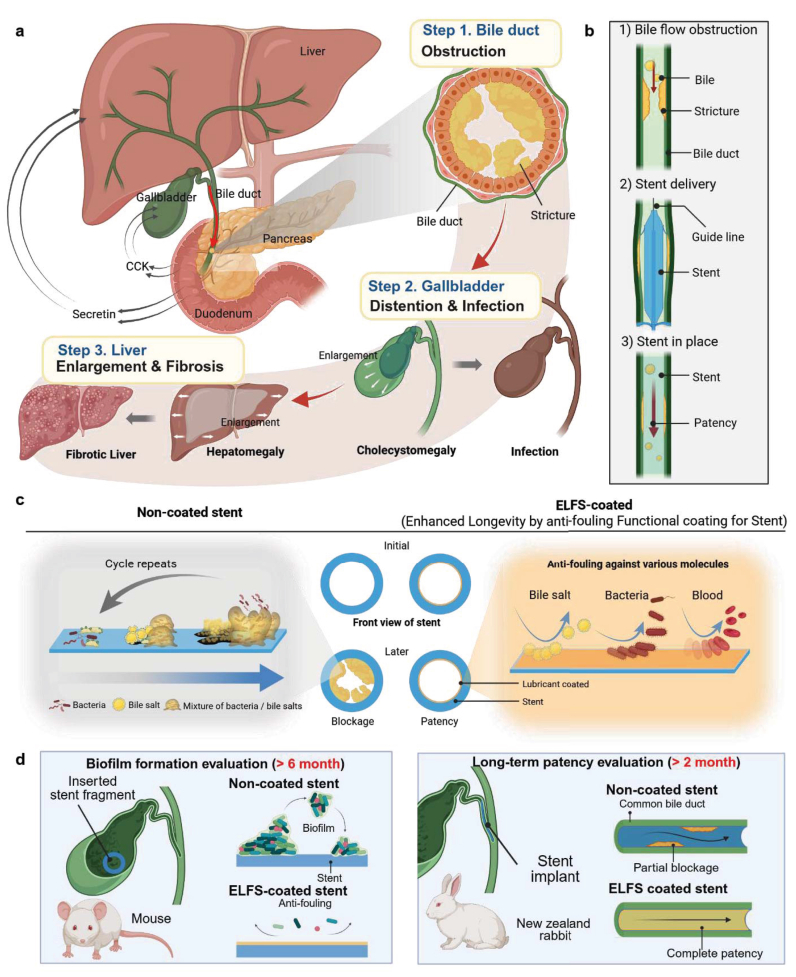
ELFS biliary-stent interface design, occlusion problems and preclinical evaluation layout.
Tae Young Kim, Won-Jong Lee, Yurim Lee et al. “Slippery dopamine–fluoropolymer hybrid surface for improving biliary stent longevity”. Figure 1. DOI: 10.1016/j.bioactmat.2026.02.003. CC BY 4.0. Figure content is unchanged.
Source figure and caption ↗ · DOI: 10.1016/j.bioactmat.2026.02.003 ↗ · CC BY 4.0 ↗
How it was evaluated
Coatings were characterized by XPS, profilometry, SEM, contact angles for bile, serum, blood and solvents, and adsorption of albumin, fibrinogen, bile, glucose, cholesterol and humic acid, with blood perfusion through polyethylene tubing and component controls (PDA only, PFP only, PFP without lubricant). Durability tests included ISO 2409 cross-cut adhesion, ultrasonication, water flushing and shear, 14-day rabbit bile flow in microchannels, thermal self-healing, swab abrasion, autoclave, ethylene oxide and gamma sterilization, and oxidative degradation. NIH 3T3 fibroblasts, human biliary epithelial cells, RAW 264.7 cells, E. coli and S. aureus were used in vitro. In vivo, intravital imaging in a mouse dorsal skinfold chamber tracked neutrophils and macrophages around coated and uncoated films up to 72 h, stent fragments were implanted in the mouse gallbladder for one week and six months, and 1 cm stents were placed in the common bile duct of rabbits (n = 4 per group) for 2 months.
Key results
The coating framework was about 300–350 nm thick, repelled liquids regardless of surface tension and showed no detectable albumin or fibrinogen adhesion, whereas a PEG coating only partly suppressed adsorption. During blood perfusion, coated polyethylene tubing remained almost stain-free, while PDA-only, PFP-only and lubricant-free PFP controls fouled fully or partly. The coating showed no detachment in the cross-cut test with ten tape peels, retained more than 80% of its lubricant after 14 days of continuous rabbit bile flow, kept nearly unchanged contact angles after more than 9 autoclave cycles, 3 ethylene oxide cycles and 3 gamma irradiation cycles, and protected the PDA layer from oxidative degradation that halved the absorbance of PDA alone within 7 days.
In vitro, the coating did not reduce fibroblast or biliary epithelial cell viability, prevented cell attachment on patterned regions, and showed virtually no E. coli or S. aureus attachment after 24 h, with no biofilm formation on coated stent fragments. In intravital imaging, neutrophils accumulated progressively around uncoated films from 3 h to 72 h, with more than 20-fold higher recruitment than around coated films at 3 h, followed by macrophages. After one week, uncoated implants showed more mast cells and macrophages, α-SMA-positive myofibroblasts and a collagen capsule, whereas coated implants showed no significant fibrotic encapsulation.
In the mouse gallbladder, uncoated stent fragments accumulated bile sludge within one week, more after six months, and bacterial colonies, while coated fragments showed markedly less sludge and no biofilm-like accumulation. Uncoated fragments were associated with about a 1.5-fold increase in ALT without histological liver changes.
In the rabbit common bile duct, contrast medium and dyed saline passed through coated stents after 2 months, whereas uncoated stents were completely occluded. Rabbits with uncoated stents showed bile leakage, a gallbladder enlarged more than seven-fold, two-fold liver enlargement, bile duct dilation (reported as over 4 times in the abstract and about seven-fold in the results), rising bilirubin from four weeks, increased ALT and AST at 2 months, periductal fibrosis, muscular hypertrophy, hepatic fibrosis and more viscous bile. Rabbits with coated stents showed no visible hepatobiliary damage and relatively stable bilirubin, ALT and AST.
Open questions
The authors note that constant flow in the bile-flow test does not replicate intermittent physiological bile flow, that FBR dynamics were imaged in dorsal skin because biliary implants are not accessible to intravital imaging, and that mouse experiments used gallbladder implantation because the mouse bile duct is too small for stents. The lubricant is gradually depleted under sustained bile shear, leaving the PFP scaffold to provide later-stage resistance, and the rabbit study was limited to 2 months for animal welfare reasons after uncoated-stent deaths in a longer pilot.
Related research
A bioinspired omniphobic surface coating on medical devices prevents thrombosis and biofouling
Blood-contacting liquid coatings provide a related interface design; ELFS examines bile ducts and immune–fibrotic responses.
Abstract-based introductionRESEARCH NEWS
Research in the news
Sources used in this summary
This summary is based on the full text and figure legends of the open-access article (PMC12924897); the supplementary data, which include several of the cited supplementary figures, were not reviewed.