Overview
SHIELD-C, from a study by researchers at the BLISS Lab (Jungmok Seo) at Yonsei University and the Department of Otorhinolaryngology-Head and Neck Surgery at Korea University Ansan Hospital with other collaborators, is a single-use snap-on cap for rigid endoscopes that replaces the solid lens surface with a liquid-liquid interface formed by a grafted polydimethylsiloxane (PDMS) brush and silicone oil. The coated cap kept transmittance above 90% across 400 to 700 nm and sliding angles below 10° for blood and other biofluids, and a single contact with an oil-loaded sponge restored visual clarity from 84.3 ± 4.8% to 98.9 ± 1.2% after mucus contamination in an ex vivo test.
Background
Endoscopic surgery depends entirely on the endoscopic image, yet the temperature and humidity difference between the body and the lens causes condensation, and blood, mucus and electrosurgical byproducts accumulate on the lens. In a cited analysis of laparoscopic cholecystectomy, the view was unobstructed for only 56% of operative time, and lens cleaning took about 7% of surgical time with 15.3 interruptions per case. Chemical anti-fog agents are short-lived and do not prevent blood or mucus adhesion, lens heating and spraying systems are difficult to fit to rigid endoscopes, and reported lubricant-infused lenses rely on silica nanoparticles, fragile micro- or nanostructures, or regeneration steps that take several minutes.
Design and approach
A PDMS brush base layer is spray-coated onto a polycarbonate (PC) cap and covalently anchored, and silicone oil is then applied as a top layer. Because PDMS and silicone oil are chemically similar, the oil swells the brush network, which acts as a reservoir: when the surface oil is depleted, diffusion from the swollen network restores the liquid-liquid interface, and an oil-loaded sponge allows external reloading during a procedure. The authors attribute the antifouling effect to the absence of fixed adsorption sites on a mobile liquid surface and the anti-fogging effect to the removal of the solid-air interface on which water nucleates. Optical simulation selected a flat PC cap with a 0.3 mm wall mounted flush against the endoscope tip to minimize back-reflection. In practice, base-coated caps are supplied ready to use and the oil layer is applied with a preloaded sponge immediately before use.
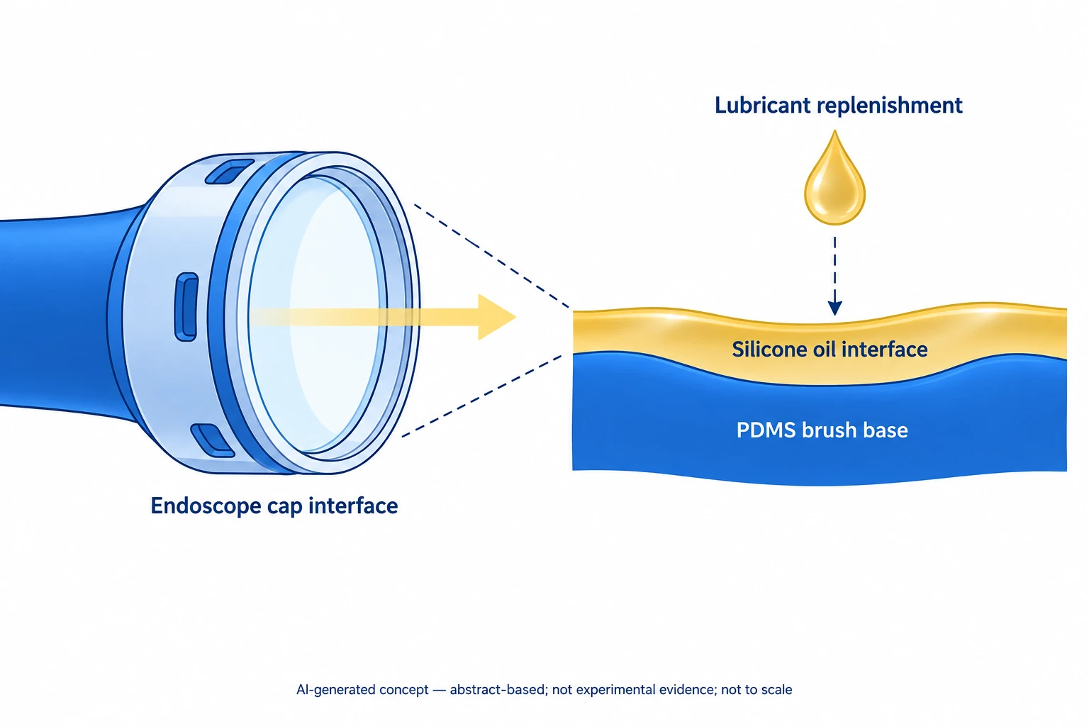
AI-generated concept based on the public abstract: SHIELD-C combines a PDMS-brush/silicone-oil interface with lubricant replenishment. The cap outline and bands represent functions, not actual geometry, experimental images or clinical outcomes.
Generated for this library to illustrate the design; it is not a figure from the paper and does not show measured data.
How it was evaluated
Coatings on PC were characterized by X-ray photoelectron spectroscopy (XPS), Fourier transform infrared spectroscopy, surface profiling, atomic force microscopy and UV-vis transmittance. Contact and sliding angles were measured for DI water, horse blood, artificial mucus, PBS, simulated urine and a 1:1 blood/mucus mixture, and stability was tested after 24 h in buffers of pH 2 to 10, orbital shaking in PBS for up to 48 h, irrigation and gauze wiping with gravimetric tracking, accelerated aging at 60 °C for 97 days and 35 kGy electron-beam sterilization. Albumin and fibrinogen adsorption were compared with bare glass and PEG-treated controls, cytocompatibility with NIH-3T3 fibroblasts, hemolysis with horse erythrocytes, and in vivo safety with SHIELD-C-coated PDMS scaffolds implanted subcutaneously in female ICR mice (n = 3). On a 4 mm rigid endoscope, SHIELD-C was compared with bare and commercial anti-fog-treated endoscopes in a 100-dip blood test, a 3 μL blood-drop test, a 38 °C/75% relative humidity fogging test with an ISO 8600-3 target, an electrocautery test with porcine tissue in a silicone tube, an ex vivo mucus refresh test and an ex vivo porcine laparoscopic model insufflated with CO2 to 12 mmHg.
Key results
The coating was about 80 nm thick with a roughness (Ra) of about 1.6 nm, and XPS showed a Si 2p peak near 102 eV that was absent on bare PC. Transmittance exceeded 90% across 400 to 700 nm, comparable to bare PC and glass, and RGB intensities of color-wheel images agreed with those of the reference substrates. Sliding angles were below 10° for all tested biofluids, and for the blood/mucus mixture the contact angle was about 100° with a sliding angle below 5°. The PDMS brush alone raised the contact angle but kept a high sliding angle, which the authors interpret as showing that droplet shedding is governed by the mobile oil layer.
Blood contact and sliding angles showed no statistically significant change after pH 2 to 10 exposure and remained unchanged after accelerated aging equivalent to about 3 years; after 35 kGy electron-beam sterilization the contact angle stayed above 100°. Irrigation removed about 0.52 mg in the first cycle and about 0.73 mg cumulatively after the second, and two gauze wipes removed about 1.69 mg, while one sponge contact transferred 1.39 mg of oil. The authors calculated that one 394.29 mg pump supports about 283 replenishments, or more than 4.5 h at one replenishment per minute.
Protein adsorption on SHIELD-C was markedly lower than on bare PC and comparable to a PEG-coated control. NIH-3T3 fibroblasts showed greater than 90% survival over 5 days, and the hemolysis ratio was below 6%, statistically comparable to the PBS control (below 7%). In mice, plasma ALT and AST remained within the healthy reference range on days 1 and 7 after implantation, and no inflammatory infiltrates were found in the heart, liver, kidney or lung.
On the endoscope, SHIELD-C kept a low blood-covered area after 100 dips in horse blood, whereas bare and commercial anti-fog-treated endoscopes became nearly fully obstructed. A blood drop slid off within 2 s and nearly 100% visibility was preserved, and in the humidity chamber SHIELD-C maintained contrast comparable to the commercial anti-fog agent. During electrocautery it prevented fogging while smoke remained visible, a single sponge refresh restored clarity from 84.3 ± 4.8% to 98.9 ± 1.2% after mucus contamination, and in the ex vivo porcine laparoscopic model it matched the commercial agent for fog control and kept a clear view under blood contamination. Friction coefficients were 0.3015 for bare PC, 0.1173 for the brush layer and 0.1409 for the complete coating.
Open questions
The authors note that the hemolysis ratio was slightly above the 5% threshold that ISO 10993-4 classifies as hemolytic, although it was statistically indistinguishable from the PBS control. Adding the silicone oil top layer slightly increased the friction coefficient relative to the brush layer alone, which the authors attribute to viscous drag of the liquid film. Occasional adhesion of mucus or luminal contents could still temporarily impair visibility and required a sponge refresh. Endoscopic performance was evaluated in benchtop and ex vivo models.
Related research
A bioinspired omniphobic surface coating on medical devices prevents thrombosis and biofouling
Both studies retain a liquid interface to resist fouling. SHIELD-C applies the idea to a replenishable cap and endoscopic visibility.
Abstract-based introductionRESEARCH NEWS
Research in the news
Sources used in this summary
This summary is based on the full main text, methods and figure legends of the published article and the text of its supporting information (figure captions S1-S16 and Tables S1-S2); Supplementary Video 1 and values plotted only in figure panels were not reviewed.