Research context and placement
Self-bonding of a shared polymer joins the conduit and flow sensor. Resealing mechanics and the antithrombotic lubricant interface are secondary connections.
Research background
Vascular sensing introduces sealing problems. This study joins sensor attachment with puncture resealing. A useful signal does not resolve leakage at a sensor attachment or needle hole. The paper therefore places sensing and conduit continuity in the same architecture while testing their functions separately.
Approach and advances
A healable, lubricated conduit integrates a related-polymer CNT sensor through self-bonding. Related polymer chemistry addresses joining the sensor to the conduit. The lubricated lumen manages blood-facing attachment, while the CNT composite converts deformation into resistance changes; these components have different roles.
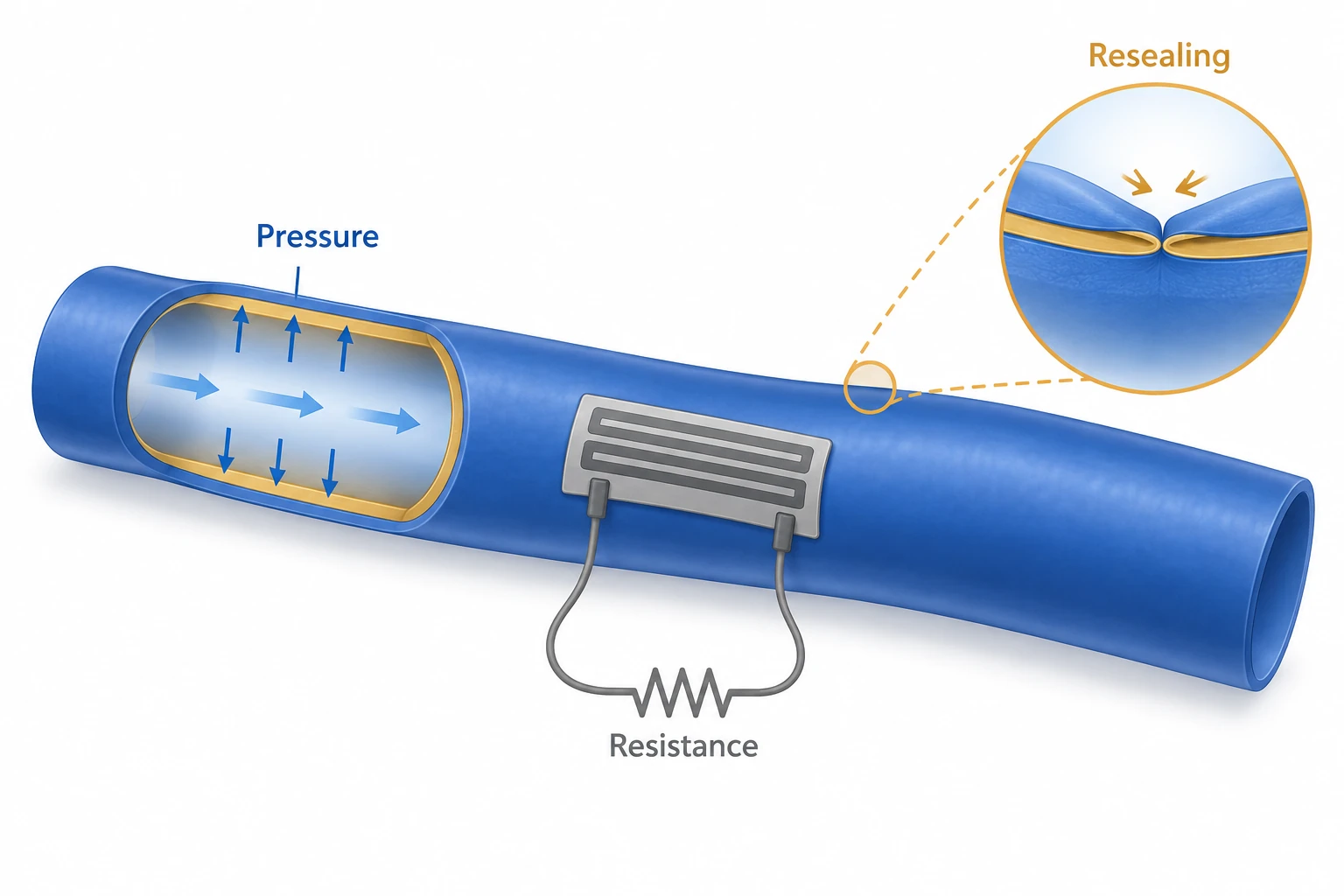
AI-generated concept separating resistance sensing of conduit pressure/deformation from resealing. The pattern, resistor symbol and seam inset are functional abstractions, not actual sensor circuitry, material cross sections, volumetric-flow accuracy or chronic vessel-replacement results.
Approved for the two functions of a benchtop integration concept; implantation, chronic patency, thrombosis prevention, sensor circuitry and demonstrated volumetric-flow measurement are excluded.
This is not a reproduced paper figure and does not establish permission to reuse the original figure.
Generated in ChatGPT on the web from concepts in a public paper and checked for conceptual consistency within the selected main-text reading and stated independent-review scope. This does not claim a complete main-text or supplement audit. Display processing is limited to WebP encoding, without cropping or content editing.
Evaluation and conditions
Sensor resistance tracked pressure fluctuations from pump-driven flow, with puncture testing for leakage and signal retention. Ex vivo porcine aorta, one-week subcutaneous implantation and three-day injured-vein wrapping test distinct endpoints. The sensor reads pressure/deformation induced by flow rather than directly counting volumetric throughput. Wrapping an injured vein differs from replacing a resected vessel segment, so the physical implantation arrangement matters more than a broad graft label.
Key findings
Leakage suppression and pressure sensing persisted after puncture, and ex vivo connections showed no reported leakage. Together these observations support retention of the tested sensing and sealing functions after local damage. Sealing an ex vivo connection is nevertheless different from prolonged thrombus-free flow in vivo, so these results do not establish chronic replacement-vessel patency.
Limits and open questions
Brief observations do not establish chronic success. Diameter, pressure, flow waveform and anticoagulation affect comparisons. Even at the same pump setting, conduit geometry and stiffness can change the pressure–resistance relationship. Clinical flow accuracy and chronic patency remain separate questions from the demonstrated short-term integration.
Related external research
Biodegradable and flexible arterial-pulse sensor for the wireless monitoring of blood flow
DOI: 10.1038/s41551-018-0336-5 ↗
Boutry describes a biodegradable wireless capacitive pulse sensor; the focal paper emphasizes a resealable conduit and sensor integration.
The external sensor is not a replacement vessel; pulse/pressure sensing and volumetric-flow calibration differ.
Abstract checkedCorresponding-author verification
Jungmok Seo: corresponding authorThis record concerns Jungmok Seo’s correspondence designation. Author order or an asterisk alone is not treated as confirmation; this check is separate from verification of the research content.
- Correspondence evidence source ↗
Corresponding Authors; Jungmok Seo; Donghee Son
BLS01_ACSNano_2023_Vascular_Graft_Blood_Flow_Sensor_MAIN.pdf; PDF page 13, AUTHOR INFORMATION → Corresponding Authors · local_public_pdf_read
- Main-text review scope
- Local published PDF pp. 10–13: sensor integration, Fig. 6, ex vivo connections, rodent in vivo methods; 3-day injured-IVC wrapping and 1-week subcutaneous observation. Additional targeted main-text reading in this pass: Local PDF Fig. 5g–m pressure/flow/puncture testing and Fig. 6 ex vivo/in vivo models; original PDF pp. 10–11 This additional reading refers to main text, not supplementary information.
- Supplementary review scope
- Supplementary information was not read. No supplementary material is listed as reviewed, and a complete SI audit is not claimed.
- Pending verification
- SI was not read. Detailed flow calibration, statistical units, chronic vessel replacement and patency require further verification. This account is limited to the reported sealing, sensor integration and short-term models.
COVERAGE & OUTREACH
Coverage and outreach
Links are checked for their relationship to this paper. Media publication does not establish independent reporting or additional experimental validation.
인공혈관을 유막 코팅? 심혈관 질환 재발 막는다 ↗
The journal-link DOI 10.1021/acsnano.2c10657 and the Seo, Son, Park, An and Kim author names identify #68.
Partial body read Read the result metadata and main body. BioIN’s posted date is June 23, while its release-date field says June 9. This is an attributed NRF republication; the NRF original itself was not opened.
혈전 생겨도 안막히는 인공혈관 소재 나왔다 ↗
The Seo–Son collaboration, ACS Nano and self-healing polymer/lubricant-coated graft match #68.
Body read Read the body, byline and date. The story attributes its source to NRF; headline wording is not expanded into direct evidence of long-term patency.
찢어지면 스스로 치료하는 인공혈관 나왔다 ↗
The references explicitly give DOI 10.1021/acsnano.2c10657 alongside Seo, Son and the self-healing graft.
Body read Read the body, date and DOI. The story relies on an NRF announcement and supplied images; its clinical survival-rate projection is not treated as experimental evidence.
인공혈관 부작용, 자가치유 소재·유막코팅 적용으로 방지 ↗
The Seo–Son collaboration and self-healing polymer/lubricant-coated graft match #68.
Partial body read Read the date and opening body identifying NRF and the study. The complete remainder of the story was not reviewed.
Sources and verification scope
Local published PDF pp. 10–13: sensor integration, Fig. 6, ex vivo connections, rodent in vivo methods; 3-day injured-IVC wrapping and 1-week subcutaneous observation. Additional targeted main-text reading in this pass: Local PDF Fig. 5g–m pressure/flow/puncture testing and Fig. 6 ex vivo/in vivo models; original PDF pp. 10–11 This additional reading refers to main text, not supplementary information.
The additional commentary is editorially approved within the stated evidence scope. This does not imply complete verification of all main-text and supplementary material.
- Additional main-text review scope
- Local published PDF pp. 10–13: sensor integration, Fig. 6, ex vivo connections, rodent in vivo methods; 3-day injured-IVC wrapping and 1-week subcutaneous observation. Additional targeted main-text reading in this pass: Local PDF Fig. 5g–m pressure/flow/puncture testing and Fig. 6 ex vivo/in vivo models; original PDF pp. 10–11 This additional reading refers to main text, not supplementary information.
- Additional supplementary review scope
- Supplementary information was not read. No supplementary material is listed as reviewed, and a complete SI audit is not claimed.
- Public publication baseline ↗ · #68 · 2026-10-03
- Crossref metadata ↗: Only public bibliographic metadata registered with Crossref was checked. This does not mean that the publisher page, abstract, or full text was read; full-text verification in the school Chrome session remains pending. license_urls lists registered links and does not establish permission to redistribute text or figures. It may include TDM or posting-policy links.
- Source for updated publication metadata ↗
Bibliographic and publication-status check; separate from main-text review. - Resealable anti-thrombotic artificial vascular graft integrated with a self-healing blood flow sensor ↗
public_bibliography · PubMed DOI/title - Resealable anti-thrombotic artificial vascular graft integrated with a self-healing blood flow sensor ↗
local_pdf_read · BLS01_ACSNano_2023_Vascular_Graft_Blood_Flow_Sensor_MAIN.pdf; pp. 10–13; Fig. 6; in vivo rodent methods; title and DOI matched; PDF not redistributed - Resealable Antithrombotic Artificial Vascular Graft Integrated with a Self-Healing Blood Flow Sensor ↗
local_pdf_read · pp. 10–13; Fig. 6; in vivo rodent methods; title and DOI matched; PDF not redistributed - Resealable Antithrombotic Artificial Vascular Graft Integrated with a Self-Healing Blood Flow Sensor ↗
local_pdf_read · Local PDF Fig. 5g–m pressure/flow/puncture testing and Fig. 6 ex vivo/in vivo models; original PDF pp. 10–11 - Resealable Antithrombotic Artificial Vascular Graft Integrated with a Self-Healing Blood Flow Sensor ↗
independent_reviewer_selected_main_text · Local published PDF pp.10–11: Fig.5g–m, ex-vivo porcine aorta, one-week subcutaneous/three-day inferior-vena-cava wrapping. - Additional commentary source ↗
Research background · BLS01_ACSNano_2023_Vascular_Graft_Blood_Flow_Sensor_MAIN.pdf; pp. 10–13; Fig. 6; in vivo rodent methods; title and DOI matched; PDF not redistributed · local_pdf_read - Additional commentary source ↗
Approach and advances · BLS01_ACSNano_2023_Vascular_Graft_Blood_Flow_Sensor_MAIN.pdf; pp. 10–13; Fig. 6; in vivo rodent methods; title and DOI matched; PDF not redistributed · local_pdf_read - Additional commentary source ↗
Limits and open questions · BLS01_ACSNano_2023_Vascular_Graft_Blood_Flow_Sensor_MAIN.pdf; pp. 10–13; Fig. 6; in vivo rodent methods; title and DOI matched; PDF not redistributed · local_pdf_read - Additional commentary source ↗
Connections to related work · Biodegradable and flexible arterial-pulse sensor for the wireless monitoring of blood flow; DOI 10.1038/s41551-018-0336-5; publisher abstract previously read; Europe PMC REST core abstractText and title/DOI/author metadata rechecked 2026-10-03; external full text not reviewed. · public_abstract_rechecked_via_EuropePMC_API - Additional commentary source ↗
Evaluation and conditions · Local PDF Fig. 5g–m pressure/flow/puncture testing and Fig. 6 ex vivo/in vivo models; original PDF pp. 10–11 · local_pdf_read - Additional commentary source ↗
Key findings · Local PDF Fig. 5g–m pressure/flow/puncture testing and Fig. 6 ex vivo/in vivo models; original PDF pp. 10–11 · local_pdf_read