Overview
This study, co-authored by Jungmok Seo (then at Brigham and Women's Hospital, Harvard Medical School, the Harvard-MIT Division of Health Sciences and Technology and the Korea Institute of Science and Technology), who now leads the BLISS Lab at Yonsei University, built a batoid-fish-inspired soft actuator in which neonatal rat cardiomyocytes on micropatterned carbon nanotube (CNT)–gelatin methacryloyl (GelMA) and poly(ethylene glycol) (PEG) hydrogels are stimulated through embedded gold (Au) microelectrodes. The optimized construct beat spontaneously at 1.0 ± 0.1 Hz on day 3 and 0.8 ± 0.1 Hz on day 9, and local stimulation tuned its beating at 0.5 and 1.0 Hz.
Background
Cardiomyocyte-driven biorobots still lack life-like movement, on-board control, control over direction, robust actuation and low-power stimulation. A soft-robotic ray reported by Parker and co-workers used optogenetic control of cardiomyocytes on PDMS, but optogenetic tools involve complicated and long development and potential toxicity at high expression levels, and rigid PDMS substrates affect the contractile behavior of cardiac cells. Flexible microelectrodes offer local electrical stimulation without genetic modification, provided they can deform with a soft body.
Design and approach
The scaffold mimics the cartilage joints and aligned fin muscles of a stingray in four layers. A micropatterned PEG hydrogel acts as the cartilage-like support, a 200 nm thick Au microelectrode with a meander pattern lies above it to tolerate deformation, a micropatterned CNT–GelMA hydrogel (5 wt% GelMA, 1 mg/mL CNT) running perpendicular to the PEG lines guides cell alignment, and neonatal rat cardiomyocytes form the top layer. Cell contraction along the CNT–GelMA lines bends the fins, and the elastic PEG pattern returns the body to its rest shape. The Au electrode is transferred to the PEG layer and enclosed by direct UV crosslinking of both hydrogels, without a sacrificial layer.
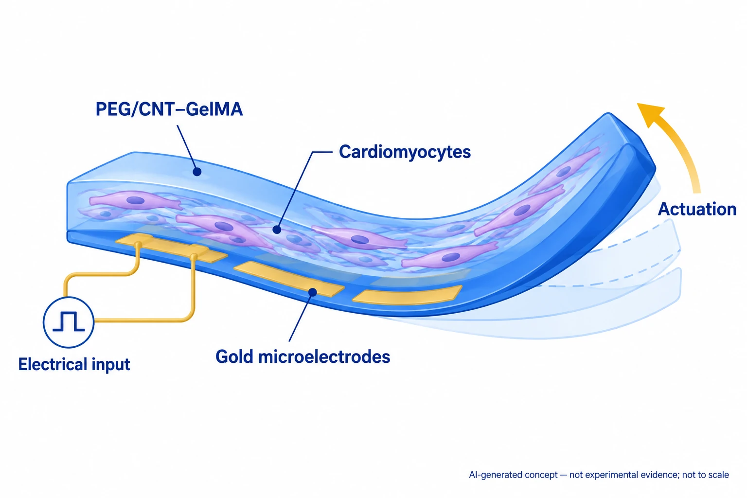
AI-generated, not-to-scale concept based on the public abstracts, showing electrically driven actuation through a PEG/CNT–GelMA structure, cardiomyocytes and gold microelectrodes.
Generated for this library to illustrate the design; it is not a figure from the paper and does not show measured data.
How it was evaluated
PEG spacings of 200, 300 and 500 µm and CNT–GelMA spacings of 50, 75 and 150 µm were compared by cell alignment, spontaneous beating rate and rolling behavior, and combined in a score index. Cardiomyocytes from 2-day-old Sprague–Dawley rats were assessed by Alamar Blue metabolic activity, sarcomeric α-actinin and connexin-43 staining, and beating rates over culture. Hydrogel moduli were measured by atomic force microscopy (AFM)-assisted nanoindentation, and the electrode by AFM roughness, resistance in culture medium at 37 °C and electrochemical impedance. Beating was paced either by external carbon rod electrodes or through the embedded Au microelectrodes at 0.5, 1.0 and 2.0 Hz, and finite element simulations compared the field distributions of the two electrode types.
Key results
CNT–GelMA lines spaced 150 µm gave the clearest alignment, but spontaneous beating was faster and more stable at 75 µm spacing (0.9 ± 0.1 Hz) than at 50 µm (0.5 ± 0.2 Hz) or 150 µm (0.2 ± 0.1 Hz). Constructs with 50 µm spacing actuated strongly but bent in the wrong direction. For the PEG support (20 wt%), 200 µm spacing prevented folding and 500 µm spacing caused irreversible rolling with weak beating; the score index was highest for 300 µm PEG and 75 µm CNT–GelMA spacing. Metabolic activity increased over 7 d of culture on scaffolds with 75 µm CNT–GelMA spacing.
On the optimized scaffold, cardiomyocytes beat spontaneously from day 3 (1.0 ± 0.1 Hz) to day 9 (0.8 ± 0.1 Hz) without a significant reduction, and their contraction detached the construct from the glass after 4 or 5 d. Young's modulus was 651 ± 326 kPa for the PEG pattern and 37 ± 16 kPa for the CNT–GelMA pattern. The fins contracted in the transverse direction while the longitudinal axis stretched, and the body returned to rest after each contraction; the construct showed self-actuating movement but not forward propulsion. With external carbon rod electrodes, beating was controlled at 0.5, 1.0 and 2.0 Hz, with smaller actuation amplitudes at higher frequencies.
The embedded electrodes had a root mean square roughness of 2.0 ± 0.4 nm and showed only slight resistance variation over 5 d in culture medium at 37 °C. Adding the CNT–GelMA layer lowered impedance at 0.1 Hz from 2.3 to 1.6 kΩ. In simulations, the microelectrodes produced a nonuniform potential with a maximum of 1 V in the central body, which the authors suggest could produce a wave-like displacement from the central body toward the fins, whereas carbon rods produced an almost uniform field and, after attenuation in the medium, delivered only 2 V of a 4 V peak to the scaffold.
Carbon rods synchronized beating at peak voltages of 0.8 to 1.5 V. Local stimulation through the Au microelectrodes required slightly higher thresholds and tuned beating at 0.5 and 1.0 Hz, but 2.0 Hz was not reached because pacing failed below 3 V, the maximum amplitude available. After 4 to 5 h of local stimulation, spontaneous beating remained around 1 Hz until day 7, similar to constructs without microelectrodes.
Open questions
The actuator did not generate forward propulsion. Local stimulation needed higher excitation voltages than external stimulation, and 2.0 Hz pacing would require amplitudes above the 3 V available. The opaque Au electrode may have partially prevented UV crosslinking of the CNT–GelMA beneath it. The Results report external pacing up to 2.0 Hz, whereas the Conclusion states control up to 1.0 Hz for both electrode types at peak voltages below 3 V.
Related research
Phototactic guidance of a tissue-engineered soft-robotic ray
DOI: 10.1126/science.aaf4292 ↗
A cardiomyocyte-driven ray with a gold skeleton uses optogenetic steering, offering a light-controlled counterpart to local electrical stimulation.
Abstract-based introductionRESEARCH NEWS
Research in the news
Sources used in this summary
This summary is based on the publisher PDF of the communication (Adv. Mater. 2018, 30, 1704189; 13 pages, local copy), including the abstract, main text, Figure 1-4 legends and the Experimental Section; the Supporting Information and Videos S1-S6 were not reviewed, and values shown only in figure panels were not used.